Japanese
English
- 有料閲覧
PPV購入案内
1,320円 (1,200円+税10%) こちらは、463ページから468ページの文献です。購入には医書.jp本会員登録が必要です。
会員登録完了後に改めて上記「購入サイトへ」を選択してください。
または「購入サイトへ」選択後に「購入ログイン」、「新規会員登録」の順に進んでください。
会員登録について詳しくはをご確認ください。
- Abstract 文献概要
- 1ページ目 Look Inside
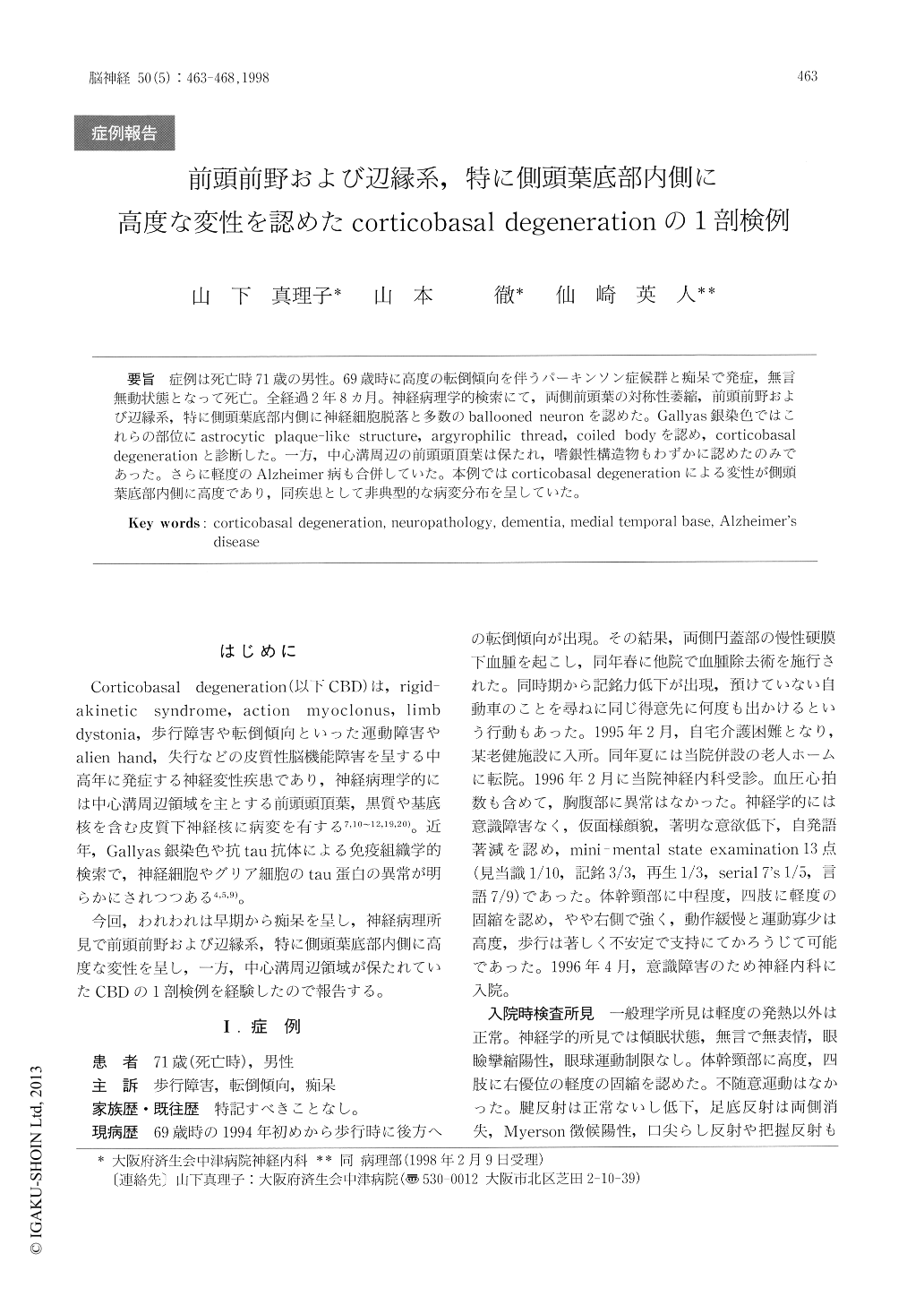
症例は死亡時71歳の男性。69歳時に高度の転倒傾向を伴うパーキンソン症候群と痴呆で発症,無言無動状態となって死亡。全経過2年8カ月。神経病理学的検索にて,両側前頭葉の対称性萎縮,前頭前野および辺縁系,特に側頭葉底部内側に神経細胞脱落と多数のballooned neuronを認めた。Gallyas銀染色ではこれらの部位にastrocytic plaque-like structure, argyrophilic thread, coiled bodyを認め,corticobasal degenerationと診断した。一方,中心溝周辺の前頭頭頂葉は保たれ,嗜銀性構造物もわずかに認めたのみであった。さらに軽度のAlzheimer病も合併していた。本例ではcorticobasal degenerationによる変性が側頭葉底部内側に高度であり,同疾患として非典型的な病変分布を呈していた。
We report a case of corticobasal degeneration (CBD) with prominent degenerative changes in the temporal base. The patient developed parkinsonism with a marked tendency to fall and dementia at the age of 69. His symptoms progressively worsened. Finally, he was akinetic and mute and died by infec-tion at 71 years of age, 32-months after the onset of his neurological symptoms. At autopsy, symmetri-cal atrophy of the prefrontal lobe was seen, yet the regions around the central sulci were preserved.
Copyright © 1998, Igaku-Shoin Ltd. All rights reserved.