Japanese
English
- 有料閲覧
- Abstract 文献概要
- 1ページ目 Look Inside
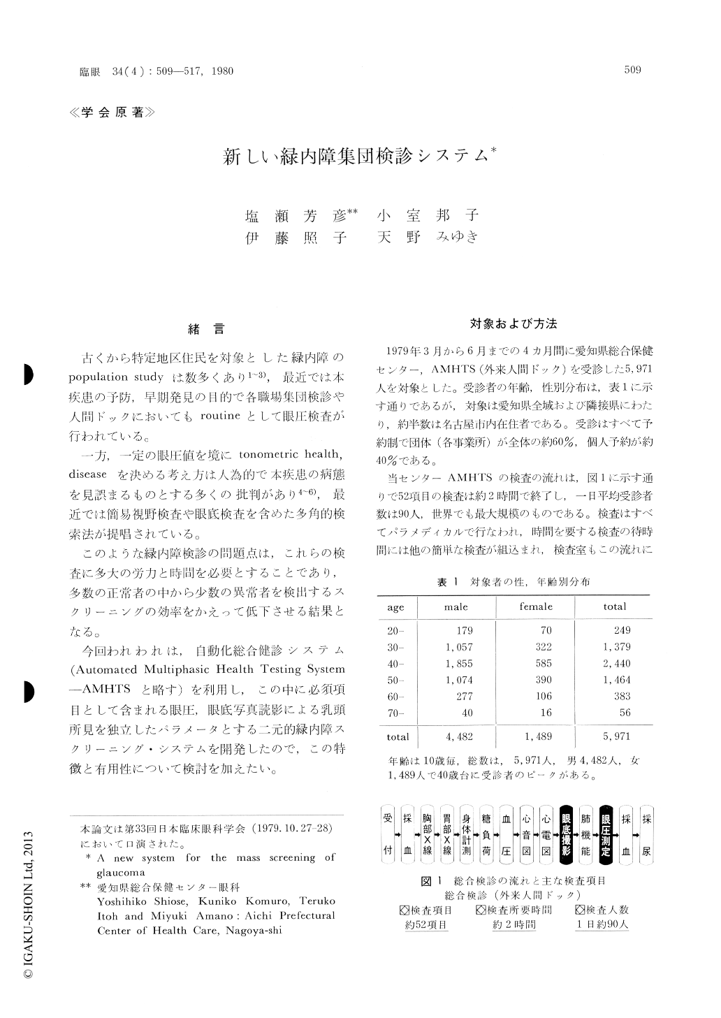
今回われわれは自動化人間ドック検査の一環として眼底写真判定を主体とした新しい緑内障集団検診システムを開発した。このシステムはパラメディカルによる一次スクリーニングと医師による二次スクリーニングからなる。
被検者は,1979年3月〜6月までに外来人間ドックを受診した5,971人である。
1)眼底写真評価法:一次スクリーニングでは乳頭量定パターン(塩瀬)を画像対比プロジェクター上で適合させ,pailorが安全域にあるものはすべて正常と判定。二次スクリーニングではCup縁,NFBD, splinter hemorrhageを参考として要精検者を出すものである。
2)眼底写真判定で40人(0,67%)の視機能異常者が発見され,その内訳は緑内障11人,低眼圧緑内障19人,初期緑内障疑10人であった。
3)眼圧スクリーニングで異常疑とされたもののうち実際視野異常あったものは,27人中4人(14.8%検出率)であったのに対し,眼底スクリーニングでは異常疑者62人中視野異常者Ib以上は30人(48.4%検出率)であった。
4)眼底スクリーニングは熟練すればパラメディカルでフィルム約90枚は,15分以内に読影可能でかつ緑内障検出率も高いことから,この方法が今後の主体となり,眼圧スクリーニングは補助的手段と考たい。
A new system for glaucoma mass-screening was designed and was put to practical trial in a group of 5,971 subjects. It was developed as a part of Automated Multiphasic Health Testing System (AMHTS). It is conducted by paramedicals inthe primary phase and is checked by specialized doctors in the secondary phase. Plane color pho-tographs and tonometry by noncontact tonometer are used for the screening of glaucoma.
In the evaluation of the optic disc as fundus photograph, four key factors are routinely assess-ed: disc pallor, cupping, nerve fiber bundle defect (NFBD) and splinter hemorrhage.

Copyright © 1980, Igaku-Shoin Ltd. All rights reserved.


