Japanese
English
- 有料閲覧
- Abstract 文献概要
- 1ページ目 Look Inside
1)広隅角緑内障13例の角膜中央部の厚さと,Specular microscopeによる角膜内皮の定量的観察を行ない細胞面積,細胞数,細胞の変形度を検討した。
角膜の厚さは0.455±0.05mm (P<0.05),細胞面積464.8±74.9μm2(P<0.01),細胞数2151.2±346.7/mm2(P<0.01)と正常に比較して有意の差を認めた。また変形度は円を1とした時の値で0.8±0.03と正常と有意の差はなかった。
2)両眼性の早期緑内障においては,進行した眼に角膜の厚さの減少と細胞の大型化を認めた。低眼圧緑内障では,眼圧が正常範囲であるにかかわらず,すでに厚さの減少と細胞の大型化を認めた。
3)広隅角緑内障において,角膜内皮は眼圧に反応し,機能的および形態的に変化することが推察された。
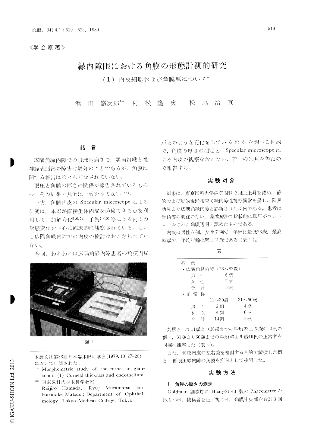
We evaluated the corneal thickness and the state of corneal endothelium using specular mi-croscope in 13 eyes with primary open angle glau-coma with well-controlled tension by medication. There was no past history of eye surgery, inflamma-tion or irritation. Twenty-four normal subjects, 14 under 30 years of age and 10 over 31 years, served as control.
The corneal thickness in glaucoma subjects averaged 0.455±0.048 mm. The value was sig-nificantly smaller than that in the control which averaged 0.505±0.076 mm.
The state of the corneal endothelium as observed by specular microscope was evaluated by image-analyzer.

Copyright © 1980, Igaku-Shoin Ltd. All rights reserved.


