Japanese
English
- 有料閲覧
PPV購入案内
1,320円 (1,200円+税10%) こちらは、879ページから882ページの文献です。購入には医書.jp本会員登録が必要です。
会員登録完了後に改めて上記「購入サイトへ」を選択してください。
または「購入サイトへ」選択後に「購入ログイン」、「新規会員登録」の順に進んでください。
会員登録について詳しくはをご確認ください。
- Abstract 文献概要
- 1ページ目 Look Inside
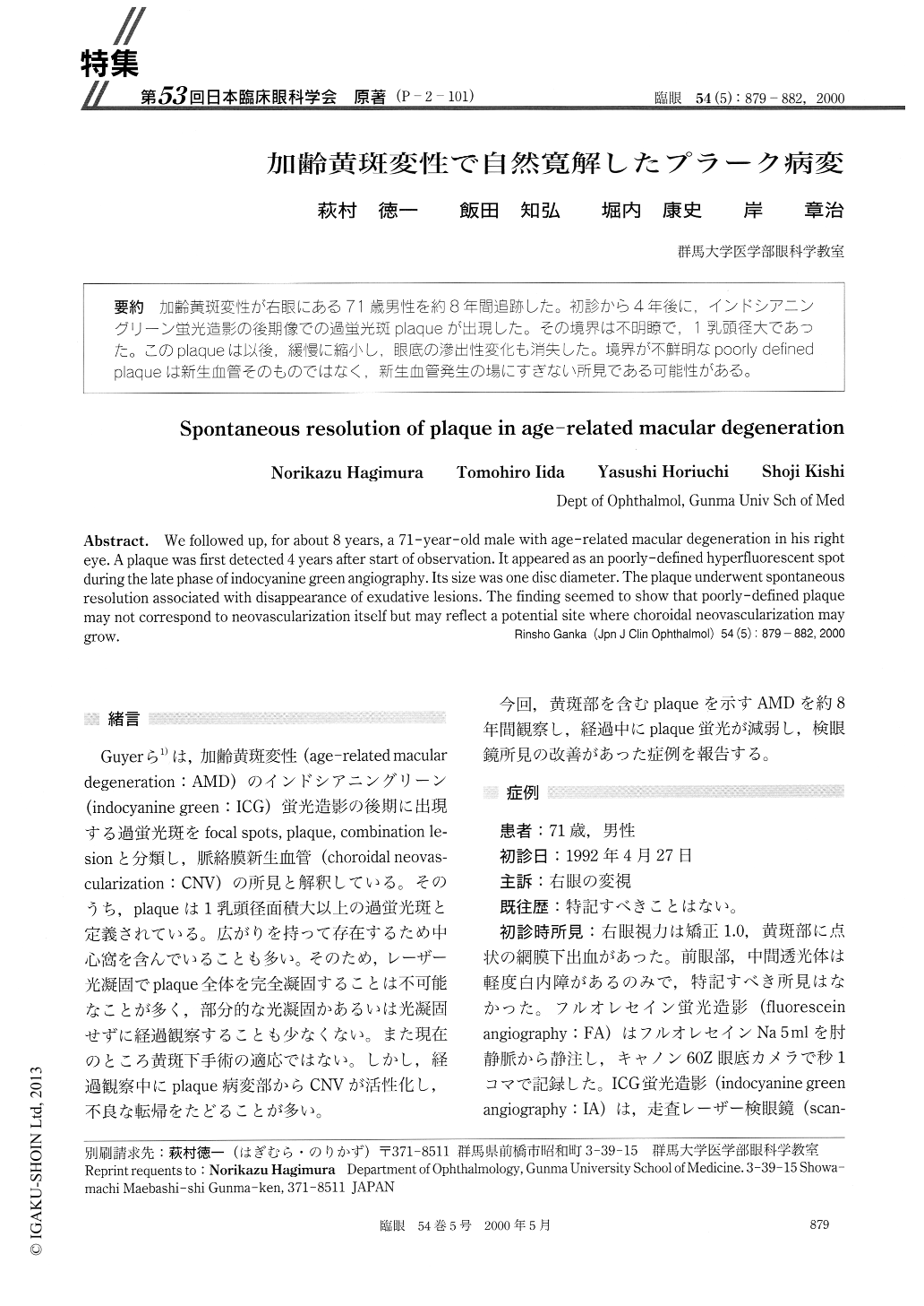
(P−2-101) 加齢黄斑変性が右眼にある71歳男性を約8年間追跡した。初診から4年後に,インドシアニングリーン蛍光造影の後期像での過蛍光斑plaqueが出現した。その境界は不明瞭で,1乳頭径大であった。このplaqueは以後,緩慢に縮小し,眼底の滲出性変化も消失した。境界が不鮮明なpoorly definedpiaqueは新生血管そのものではなく,新生血管発生の場にすぎない所見である可能性がある。
We followed up, for about 8 years, a 71-year-old male with age-related macular degeneration in his right eye. A plaque was first detected 4 years after start of observation. It appeared as an poorly-defined hyperfluorescent spot during the late phase of indocyanine green angiography. Its size was one disc diameter. The plaque underwent spontaneous resolution associated with disappearance of exudative lesions. The finding seemed to show that poorly-defined plaque may not correspond to neovascularization itself but may reflect a potential site where choroidal neovascularization may grow.
Copyright © 2000, Igaku-Shoin Ltd. All rights reserved.