Japanese
English
- 有料閲覧
PPV購入案内
1,320円 (1,200円+税10%) こちらは、751ページから756ページの文献です。購入には医書.jp本会員登録が必要です。
会員登録完了後に改めて上記「購入サイトへ」を選択してください。
または「購入サイトへ」選択後に「購入ログイン」、「新規会員登録」の順に進んでください。
会員登録について詳しくはをご確認ください。
- Abstract 文献概要
- 1ページ目 Look Inside
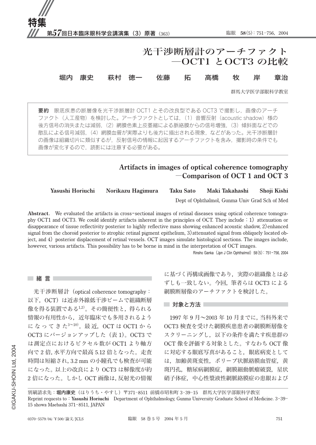
眼底疾患の断層像を光干渉断層計OCT1とその改良型であるOCT3で撮影し,画像のアーチファクト(人工産物)を検討した。アーチファクトとしては,(1)音響反射(acoustic shadow)様の後方信号の消失または減弱,(2)網膜色素上皮萎縮による脈絡膜からの信号増強,(3)傾斜面などでの散乱による信号減弱,(4)網膜血管が実際よりも後方に描出される現象,などがあった。光干渉断層計の画像は組織切片に類似するが,反射信号の情報に起因するアーチファクトを含み,撮影時の条件でも画像が変化するので,読影には注意する必要がある。
We evaluated the artifacts in cross-sectional images of retinal diseases using optical coherence tomography OCT1 and OCT3. We could identify artifacts inherent in the principles of OCT. They include:1)attenuation or disappearance of tissue reflectivity posterior to highly reflective mass showing enhanced acoustic shadow,2)enhanced signal from the choroid posterior to atrophic retinal pigment epithelium,3)attenuated signal from obliquely located object,and 4)posterior displacement of retinal vessels. OCT images simulate histological sections. The images include,however,various artifacts. This possibility has to be borne in mind in the interpretation of OCT images.
Copyright © 2004, Igaku-Shoin Ltd. All rights reserved.