Japanese
English
- 有料閲覧
PPV購入案内
1,320円 (1,200円+税10%) こちらは、905ページから908ページの文献です。購入には医書.jp本会員登録が必要です。
会員登録完了後に改めて上記「購入サイトへ」を選択してください。
または「購入サイトへ」選択後に「購入ログイン」、「新規会員登録」の順に進んでください。
会員登録について詳しくはをご確認ください。
- Abstract 文献概要
- 1ページ目 Look Inside
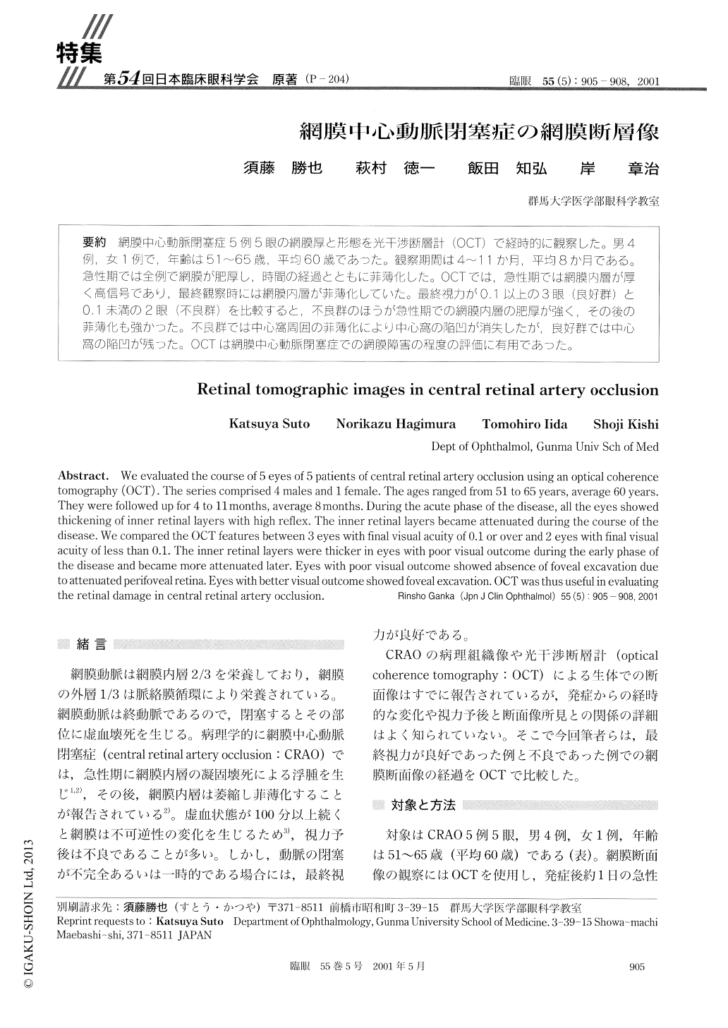
網膜中心動脈閉塞症5例5眼の網膜厚と形態を光干渉断層計(OCT)で経時的に観察した。男4例,女1例で,年齢は51〜65歳,平均60歳であった。観察期間は4〜11か月,平均8か月である。急性期では全例で網膜が肥厚し,時間の経過とともに菲薄化した。OCTでは,急性期では網膜内層が厚く高信号であり,最終観察時には網膜内層が菲薄化していた。最終視力が0.1以上の3眼(良好群)と0.1未満の2眼(不良群)を比較すると,不良群のほうが急性期での網膜内層の肥厚が強く,その後の菲薄化も強かった。不良群では中心窩周囲の菲薄化により中心窩の陥凹が消失したが,良好群では中心窩の陥凹が残った。OCTは網膜中心勤脈閉塞症での網膜障害の程度の評価に有用であった。
We evaluated the course of 5 eyes of 5 patients of central retinal artery occlusion using an optical coherencetomography (OCT) . The series comprised 4 males and 1 female. The ages ranged from 51 to 65 years, average 60 years.They were followed up for 4 to 11 months, average 8 months. During the acute phase of the disease, all the eyes showedthickening of inner retinal layers with high reflex. The inner retinal layers became attenuated during the course of thedisease. We compared the OCT features between 3 eyes with final visual acuity of 0.1 or over and 2 eyes with final visualacuity of less than 0.1. The inner retinal layers were thicker in eyes with poor visual outcome during the early phase ofthe disease and became more attenuated later. Eyes with poor visual outcome showed absence of foveal excavation dueto attenuated perifoveal retina. Eyes with better visual outcome showed foveal excavation. OCT was thus useful in evaluatingthe retinal damage in central retinal artery occlusion.
Copyright © 2001, Igaku-Shoin Ltd. All rights reserved.