Japanese
English
- 有料閲覧
- Abstract 文献概要
- 1ページ目 Look Inside
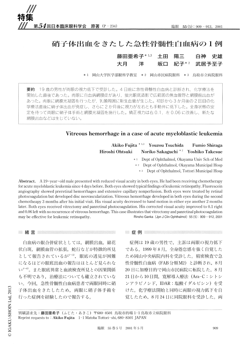
19歳の男性が両眼の視力低下で受診した。4日前に急性骨髄性白血病と診断され,化学療法を開始した直後であった。両眼に白血病網膜症があり,蛍光眼底造影で広範囲の無血管野と網膜前出血があった。両眼に網膜光凝固を行ったが,乳頭周囲に新生血管が生じた。初診から3か月後の2回目の化学療法直後に硝子体出血が発症し,さらに2か月後に視力が左右とも手動弁に低下した。全身状態の安定を待って両眼に硝子体手術と網膜光凝固を施行した。矯正視力は右0.1,左0.06に改善し,新たな網膜出血などは生じていない。
A 19-year-old male presented with reduced visual acuity in both eyes. He had been receiving chemotherapyfor acute myeloblastic leukemia since 4 days before. Both eyes showed typical findings of leukemic retinopathy.Fluorescein angiography showed preretinal hemorrhages and extensive capillary nonperfusion. Both eyes were treated by retinalphotocoagulation but developed disc neovascularization. Vitreous hemorrhage developed in both eyes during the secondchemotheapy 3 months after his initial visit. His visual acuity decreased to hand motion in either eye another 2 monthslater. Both eyes received vitrectomy and panretinal photocoagulation. His corrected visual acuity improved to 0.1 rightand 0.06 left with no recurrence of vitreous hemorrhage. This case illustrates that vitrectomy and panretinal photocoagulationmay be effective for leukemic retinopathy.

Copyright © 2001, Igaku-Shoin Ltd. All rights reserved.


