Japanese
English
- 有料閲覧
- Abstract 文献概要
- 1ページ目 Look Inside
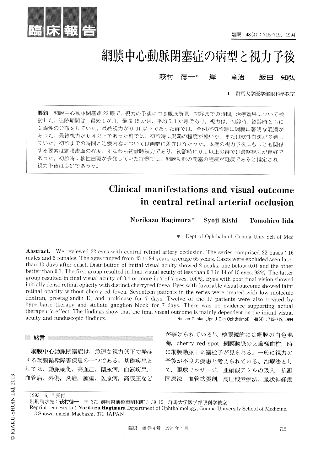
網膜中心動脈閉塞症22眼で,視力の予後につき眼底所見,初診までの時間,治療効果について検討した。追跡期間は,最短1か月,最長15か月,平均5.1か月であり,視力は,初診時,終診時ともに2峰性の分布をしていた。最終視力が0.01以下であった群では,全例が初診時に網膜に著明な混濁があった。最終視力が0.4以上であった群では,初診時に混濁の程度が軽いか,または軟性白斑が多発していた。初診までの時間と治療内容については両群に差異はなかった。本症の視力予後にもっとも関係する要素は網膜虚血の程度,すなわち初診時視力であり,初診時に0.1以上の群では最終視力が良好であった。初診時に軟性白斑が多発していた症例では,網膜動脈の閉塞の程度が軽度であると推定され,視力予後は良好であった。
We reviewed 22 eyes with central retinal artery occlusion. The series comprised 22 cases 16 males and 6 females. The ages ranged from 45 to 84 years, average 65 years. Cases were excluded seen later than 10 days after onset. Distribution of initial visual acuity showed 2 peaks, one below 0.01 and the other better than 0.1. The first group resulted in final visual acuity of less than 0.1 in 14 of 15 eyes, 93%. The latter group resulted in final visual acuity of 0.4 or more in 7 of 7 eyes, 100%. Eyes with poor final vision showed initially dense retinal opacity with distinct cherryred fovea. Eyes with favorable visual outcome showed faint retinal opacity without cherryred fovea. Seventeen patients in the series were treated with low molecule dextran, prostaglandin E1 and urokinase for 7 days. Twelve of the 17 patients were also treated by hyperbaric therapy and stellate ganglion block for 7 days. There was no evidence supporting actual therapeutic effect. The findings show that the final visual outcome is mainly dependent on the initial visual acuity and funduscopic findings.

Copyright © 1994, Igaku-Shoin Ltd. All rights reserved.


