Japanese
English
- 有料閲覧
- Abstract 文献概要
- 1ページ目 Look Inside
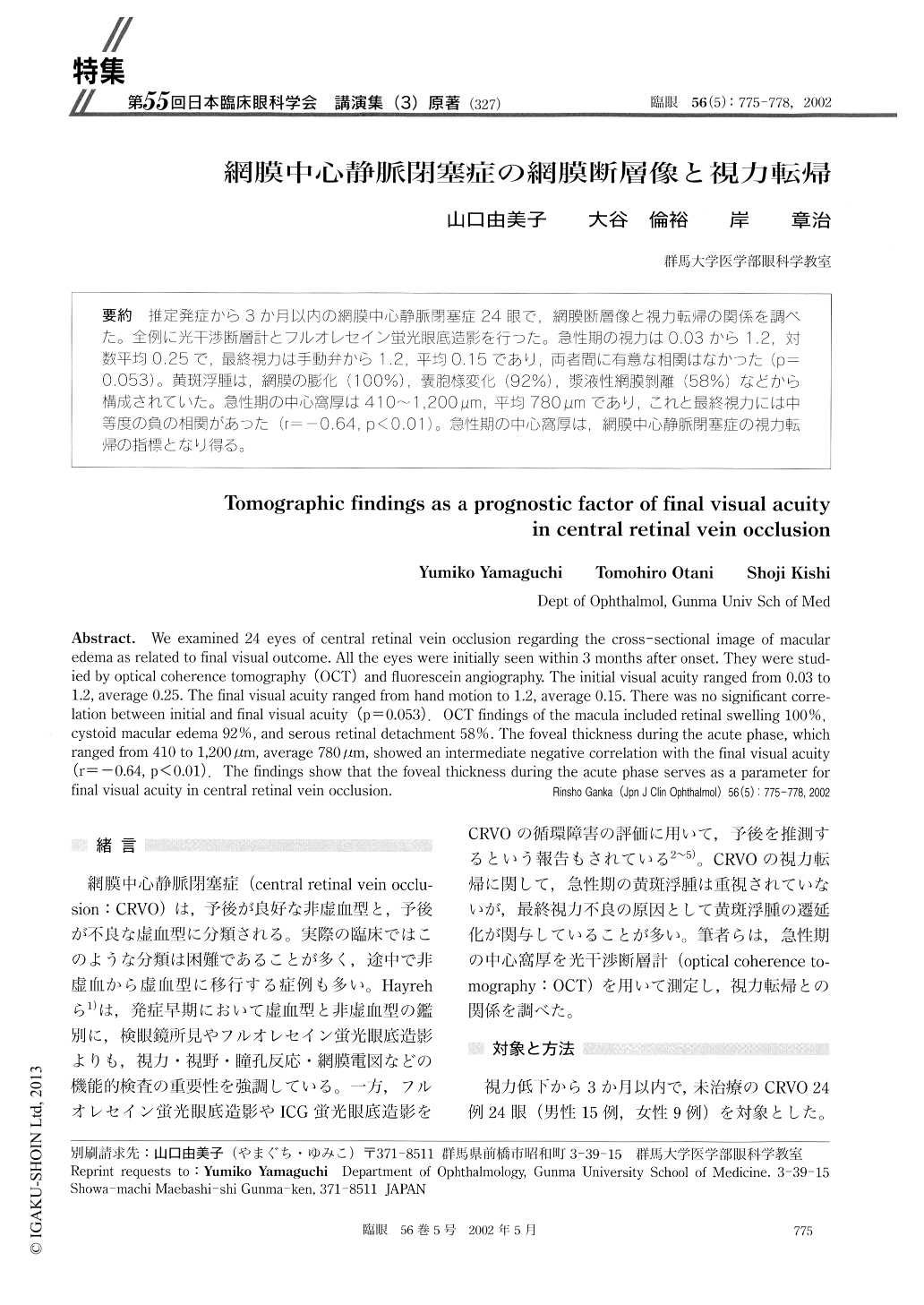
推定発症から3か月以内の網膜中心静脈閉塞症24眼で,網膜断層像と視力転帰の関係を調べた。全例に光干渉断層計とフルオレセイン蛍光眼底造影を行った。急性期の視力は0.03から1.2,対数平均0.25で,最終視力は手動弁から1.2,平均0.15であり,両者間に有意な相関はなかった(p=0.053)。黄斑浮腫は,網膜の膨化(100%),嚢胞様変化(92%),漿液性網膜剥離(58%)などから構成されていた。急性期の中心窩厚は410〜1.200μm,平均780μmであり,これと最終視力には中等度の負の相関があった(r=−0.64,p<0.01)。急性期の中心窩厚は,網膜中心静脈閉塞症の視力転帰の指標となり得る。
We examined 24 eyes of central retinal vein occlusion regarding the cross-sectional image of macular edema as related to final visual outcome. All the eyes were initially seen within 3 months after onset. They were stud-ied by optical coherence tomography (OCT) and fluorescein angiography. The initial visual acuity ranged from 0.03 to 1.2, average 0.25. The final visual acuity ranged from hand motion to 1.2, average 0.15. There was no significant corre-lation between initial and final visual acuity (p=0.053). OCT findings of the macula included retinal swelling 100%, cystoid macular edema 92%, and serous retinal detachment 58%. The foveal thickness during the acute phase, which ranged from 410 to 1,200lμm, average 780 μm, showed an intermediate negative correlation with the final visual acuity (r=-0.64, p< 0.01). The findings show that the foveal thickness during the acute phase serves as a parameter for final visual acuity in central retinal vein occlusion.

Copyright © 2002, Igaku-Shoin Ltd. All rights reserved.


