Japanese
English
- 有料閲覧
PPV購入案内
1,320円 (1,200円+税10%) こちらは、385ページから391ページの文献です。購入には医書.jp本会員登録が必要です。
会員登録完了後に改めて上記「購入サイトへ」を選択してください。
または「購入サイトへ」選択後に「購入ログイン」、「新規会員登録」の順に進んでください。
会員登録について詳しくはをご確認ください。
- Abstract 文献概要
- 1ページ目 Look Inside
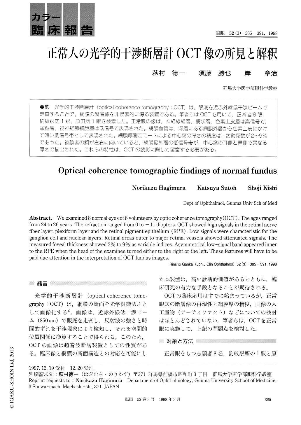
光学的干渉断層計(optical coherence tomography:OCT)は可眼底を近赤外線低干渉ビームで走査することで,網膜の断層像を非侵襲的に得る装置である。筆者らはOCTを用いて,正常者8眼,豹紋眼底1眼,原田病1眼を検索した。正常眼の像は,神経線維層,網状層,色素上皮層は高信号で,顆粒層,視神経節細胞層は低信号で表現された。網膜血管は,深層にある網膜外層から色素上皮にかけて暗い低信号帯として表現された。網膜厚測定モードによる中心窩の厚さの精度は,変動係数が2〜9%であった。被験者の顔が左右に向いていると,網膜最外層の低信号帯が,中心窩の耳側と鼻側で異なる厚さで描出された。これらの特性は,OCTの読影に際して留意する必要がある。
We examined 8 normal eyes of 8 volunteers by optic coherence tomography (OCT) . The ages ranged from 24 to 26 years. The refraction ranged from 0 to -11 diopters. OCT showed high signals in the retinal nerve fiber layer, plexiform layer and the retinal pigment epithelium (RPE) . Low signals were characteristic for the ganglion cell and nuclear layers. Retinal areas outer to major retinal vessels showed attenuated signals. The measured foveal thickness showed 2% to 9% as variable indices. Asymmetrical low-signal band appeared inner to the RPE when the head of the examinee turned either to the right or the left. These features will have to be paid due attention in the interpretation of OCT fundus images.
Copyright © 1998, Igaku-Shoin Ltd. All rights reserved.