Japanese
English
- 有料閲覧
PPV購入案内
1,320円 (1,200円+税10%) こちらは、341ページから344ページの文献です。購入には医書.jp本会員登録が必要です。
会員登録完了後に改めて上記「購入サイトへ」を選択してください。
または「購入サイトへ」選択後に「購入ログイン」、「新規会員登録」の順に進んでください。
会員登録について詳しくはをご確認ください。
- Abstract 文献概要
- 1ページ目 Look Inside
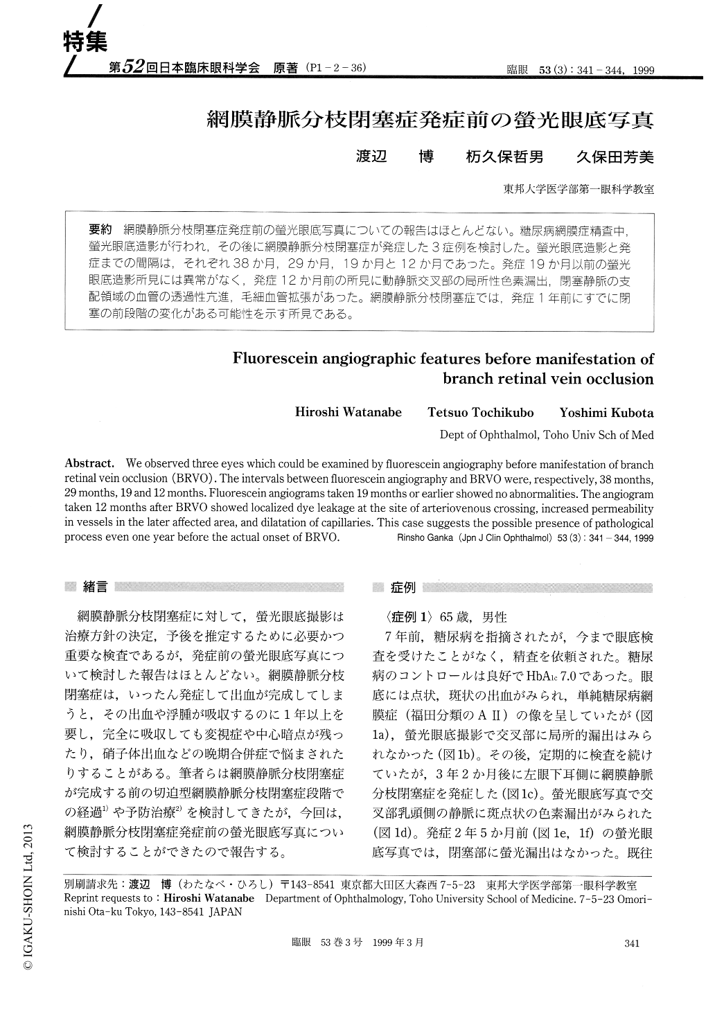
(P1-2-36) 網膜静脈分枝閉塞症発症前の螢光眼底写真についての報告はほとんどない。糖尿病網膜症精査中,螢光眼底造影が行われ,その後に網膜静脈分枝閉塞症が発症した3症例を検討した。螢光眼底造影と発症までの間隔は,それぞれ38か月,29か月,19か月と12か月であった。発症19か月以前の螢光眼底造影所見には異常がなく,発症12か月前の所見に動静脈交叉部の局所性色素漏出,閉塞静脈の支配領域の血管の透過性亢進,毛細血管拡張があった。網膜静脈分枝閉塞症では,発症1年前にすでに閉塞の前段階の変化がある可能性を示す所見である。
We observed three eyes which could be examined by fluorescein angiography before manifestation of branch retinal vein occlusion (BRVO). The intervals between fluorescein angiography and BRVO were, respectively, 38 months, 29 months, 19 and 12 months. Fluorescein angiograms taken 19 months or earlier showed no abnormalities. The angiogram taken 12 months after BRVO showed localized dye leakage at the site of arteriovenous crossing, increased permeability in vessels in the later affected area, and dilatation of capillaries. This case suggests the possible presence of pathological process even one year before the actual onset of BRVO.
Copyright © 1999, Igaku-Shoin Ltd. All rights reserved.