Japanese
English
臨床実験
螢光眼底写真による網膜静脈閉塞症の検討
Fluorescein Studies on Occlusion of the Retinal Vein
谷 道之
1
Michiyuki Tani
1
1京都府立医科大学眼科学教室
1Department of Ophthalmology, Kyoto Prefectural University of Medicine
pp.1365-1371
発行日 1967年12月15日
Published Date 1967/12/15
DOI https://doi.org/10.11477/mf.1410203766
- 有料閲覧
- Abstract 文献概要
- 1ページ目 Look Inside
I.緒言
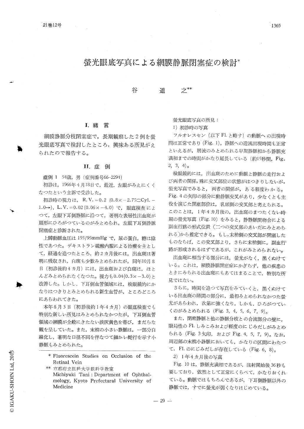
網膜静脈分枝閉塞症で,長期観察した2例を螢光眼底写真で検討したところ,興味ある所見がえられたので報告する。
Two cases with occlusion of the retinal veia were examined by fluorescein fundus photo-graphy and were followed up for a period of more than one year.
In the fresh stage, the site of the occlusion and lag of the venous flow were clearly demo-nstrated by this method. The fluorescein pho-tograph revealed remarkable and diffuse lea-kages of fluorescein between hemorrhagic fle-cks of the retina in the territory of the occluded vein. It is most likely that retinal hemorrhages occur through capillary beds in this disease.

Copyright © 1967, Igaku-Shoin Ltd. All rights reserved.


