Japanese
English
- 有料閲覧
PPV購入案内
1,320円 (1,200円+税10%) こちらは、1889ページから1892ページの文献です。購入には医書.jp本会員登録が必要です。
会員登録完了後に改めて上記「購入サイトへ」を選択してください。
または「購入サイトへ」選択後に「購入ログイン」、「新規会員登録」の順に進んでください。
会員登録について詳しくはをご確認ください。
- Abstract 文献概要
- 1ページ目 Look Inside
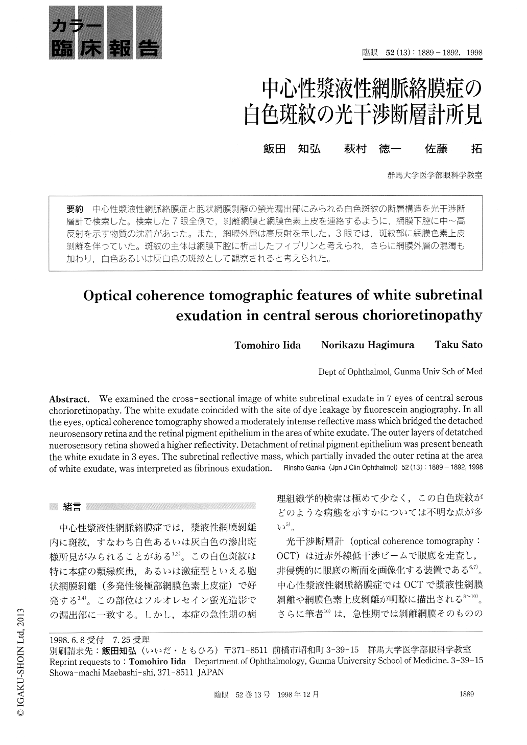
中心性漿液性網脈絡膜症と胞状網膜剥離の螢光漏出部にみられる白色斑紋の断層構造を光干渉断層計で検索した。検索した7眼全例で,剥離網膜と網膜色素上皮を連絡するように回網膜下腔に中〜高反射を示す物質の沈着があった。また時網膜外層は高反射を示した。3眼では,斑紋部に網膜色素上皮剥離を伴っていた。斑紋の主体は網膜下腔に析出したフィブリンと考えられ,さらに網膜外層の混濁も加わり,白色あるいは灰白色の斑紋として観察されると考えられた。
We examined the cross- sectional image of white subretinal exudate in 7 eyes of central serous chorioretinopathy. The white exudate coincided with the site of dye leakage by fluorescein angiography. In all the eyes, optical coherence tomography showed a moderately intense reflective mass which bridged the detached neurosensory retina and the retinal pigment epithelium in the area of white exudate. The outer layers of detatched nuerosensory retina showed a higher reflectivity. Detachment of retinal pigment epithelium was present beneath the white exudate in 3 eyes. The subretinal reflective mass, which partially invaded the outer retina at the area of white exudate, was interpreted as fibrinous exudation.
Copyright © 1998, Igaku-Shoin Ltd. All rights reserved.