Japanese
English
- 有料閲覧
PPV購入案内
1,320円 (1,200円+税10%) こちらは、1519ページから1521ページの文献です。購入には医書.jp本会員登録が必要です。
会員登録完了後に改めて上記「購入サイトへ」を選択してください。
または「購入サイトへ」選択後に「購入ログイン」、「新規会員登録」の順に進んでください。
会員登録について詳しくはをご確認ください。
- Abstract 文献概要
- 1ページ目 Look Inside
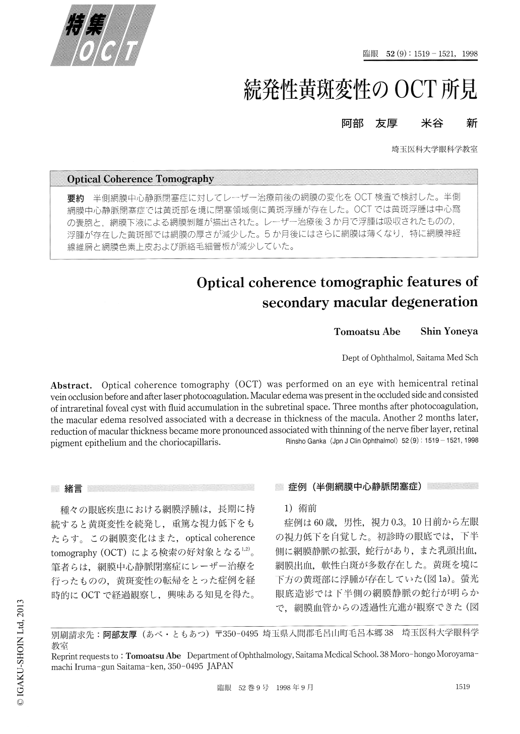
半側網膜中心静脈閉塞症に対してレーザー治療前後の網膜の変化をOCT検査で検討した。半側網膜中心静脈閉塞症では黄斑部を境に閉塞領域側に黄斑浮腫が存在した。OCTでは黄斑浮腫は中心窩の嚢胞と,網膜下液による網膜剥離が描出された。レーザー治療後3か月で浮腫は吸収されたものの,浮腫が存在した黄斑部では網膜の厚さが減少した。5か月後にはさらに網膜は薄くなり,特に網膜神経線維層と網膜色素上皮および脈絡毛細管板が減少していた。
Optical coherence tomography (OCT) was performed on an eye with hemicentral retinal vein occlusion before and after laser photocoagulation. Macular edema was present in the occluded side and consisted of intraretinal foveal cyst with fluid accumulation in the subretinal space. Three months after photocoagulation, the macular edema resolved associated with a decrease in thickness of the macula. Another 2 months later, reduction of macular thickness became more pronounced associated with thinning of the nerve fiber layer, retinal pigment epithelium and the choriocapillaris.
Copyright © 1998, Igaku-Shoin Ltd. All rights reserved.