Japanese
English
- 有料閲覧
PPV購入案内
1,320円 (1,200円+税10%) こちらは、1501ページから1509ページの文献です。購入には医書.jp本会員登録が必要です。
会員登録完了後に改めて上記「購入サイトへ」を選択してください。
または「購入サイトへ」選択後に「購入ログイン」、「新規会員登録」の順に進んでください。
会員登録について詳しくはをご確認ください。
- Abstract 文献概要
- 1ページ目 Look Inside
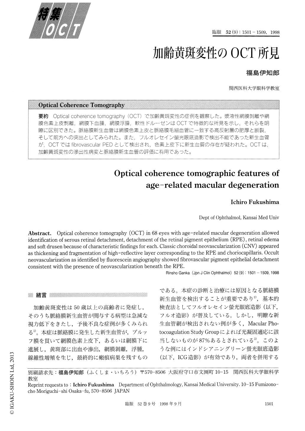
Optical coherence tomography (OCT)で加齢黄斑変性の症例を観察した。漿液性網膜剥離や網膜色素上皮剥離,網膜下血腫、網膜浮腫,軟性ドルーゼンはOCTで特徴的な所見を示し,それらを明瞭に区別できた。脈絡膜新生血管は網膜色素上皮と脈絡膜毛細血管に一致する高反射層の肥厚と断裂,そして前方への突出としてみられた。また,フルオレセイン螢光眼底造影で検出不能であった新生血管が,OCTではfibrovascular PEDとして検出され,色素上皮下に新生血管の存在が疑われた。OCTは,加齢黄斑変性の滲出性病変と脈絡膜新生血管の評価に有用であった。
Optical coherence tomography (OCT) in 68 eyes with age-related macular degeneration allowed identification of serous retinal detachment, detachment of the retinal pigment epithelium (RPE) , retinal edema and soft drusen because of characteristic findings for each. Classic choroidal neovascularization (CNV) appeared as thickening and fragmentation of high-reflective layer corresponding to the RPE and choriocapillaris. Occult neovascularization as identified by fluorescein angiography showed fibrovascular pigment epithelial detachment consistent with the presence of neovascularization beneath the RPE.
Copyright © 1998, Igaku-Shoin Ltd. All rights reserved.