Japanese
English
- 有料閲覧
PPV購入案内
1,320円 (1,200円+税10%) こちらは、1109ページから1112ページの文献です。購入には医書.jp本会員登録が必要です。
会員登録完了後に改めて上記「購入サイトへ」を選択してください。
または「購入サイトへ」選択後に「購入ログイン」、「新規会員登録」の順に進んでください。
会員登録について詳しくはをご確認ください。
- Abstract 文献概要
- 1ページ目 Look Inside
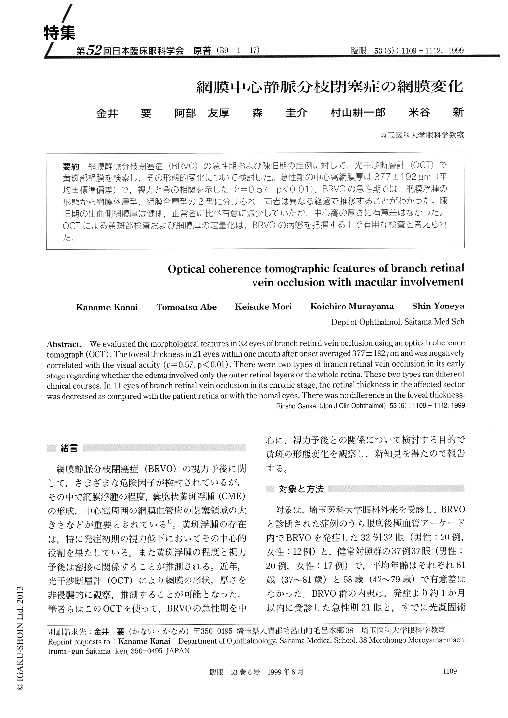
(B9-1-17) 網膜静脈分枝閉塞症(BRVO)の急性期および陳旧期の症例に対して,光干渉断層計(OCT)で黄斑部網膜を検索し,その形態的変化について検討した。急性期の中心窩網膜厚は377±192μm(平均±標準偏差)で,視力と負の相関を示した(r=0.57,p<0.01)。BRVOの急性期では,網膜浮腫の形態から網膜外層型,網膜全層型の2型に分けられ,両者は異なる経過で推移することがわかった。陳旧期の出血側網膜厚は健側,正常者に比べ有意に減少していたが,中心窩の厚さに有意差はなかった。OCTによる黄斑部検査および網膜厚の定量化は,BRVOの病態を把握ずる上で有用な検査と考えられた。
We evaluated the morphological features in 32 eyes of branch retinal vein occlusion using an optical coherence tomograph (OCT) . The foveal thickness in 21 eyes within one month after onset averaged 377 ± 192 jim and was negatively correlated with the visual acuity (r = 0.57, p < 0.01) . There were two types of branch retinal vein occlusion in its early stage regarding whether the edema involved only the outer retinal layers or the whole retina. These two types ran different clinical courses. In 11 eyes of branch retinal vein occlusion in its chronic stage, the retinal thickness in the affected sector was decreased as compared with the patient retina or with the nomal eyes. There was no difference in the foveal thickness.
Copyright © 1999, Igaku-Shoin Ltd. All rights reserved.