Japanese
English
- 有料閲覧
PPV購入案内
1,320円 (1,200円+税10%) こちらは、1619ページから1624ページの文献です。購入には医書.jp本会員登録が必要です。
会員登録完了後に改めて上記「購入サイトへ」を選択してください。
または「購入サイトへ」選択後に「購入ログイン」、「新規会員登録」の順に進んでください。
会員登録について詳しくはをご確認ください。
- Abstract 文献概要
- 1ページ目 Look Inside
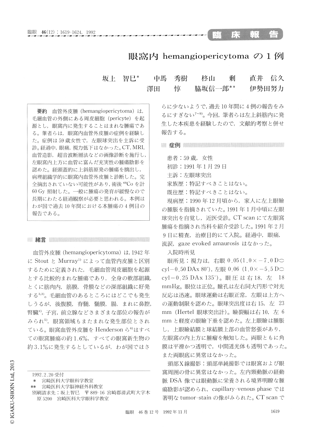
血管外皮腫(hemangiopericytoma)は,毛細血管の外側にある周皮細胞(pericyte)を起源とし,眼窩内に発生することはまれな腫瘍である。筆者らは,眼窩内血管外皮腫の症例を経験した。症例は59歳女性で,左眼球突出を主訴に受診。経過中,眼痛,視力低下はなかった。CT,MRI,血管造影,超音波断層法などの画像診断を施行し,左眼窩内上方に血管に富んだ充実性の腫瘍陰影を認めた。経頭蓋的に上斜筋原発の腫瘍を摘出し,病理組織学的に眼窩内血管外皮腫と診断した。完全摘出されていない可能性があり,術後60Coを計60Gy照射した。一般に腫瘍の発育が緩慢なので長期にわたる経過観察が必要と思われる。本例はわが国で過去10年間における本腫瘍の4例目の報告である。
A 59-year-old woman presented with progressive proptosis of her left eye of one month's duration. She was free of other symptoms as pain, visual loss or diplopia. A well-circumscribed and vascularized mass was detected in the superior nasal portion ofthe left orbit by computed tomography, magnetic resonance imaging, X-ray angiography and ultrasonography. The orbital mass was surgically removed. Histopathological studies showed a 'staghorn appearance' which is the characteristic vascular pattern of hemangiopericytoma. The pre-sent case is the fourth reported one of hemangio-pericytoma in Japan.
Copyright © 1992, Igaku-Shoin Ltd. All rights reserved.