Japanese
English
- 有料閲覧
PPV購入案内
1,320円 (1,200円+税10%) こちらは、1047ページから1049ページの文献です。購入には医書.jp本会員登録が必要です。
会員登録完了後に改めて上記「購入サイトへ」を選択してください。
または「購入サイトへ」選択後に「購入ログイン」、「新規会員登録」の順に進んでください。
会員登録について詳しくはをご確認ください。
- Abstract 文献概要
- 1ページ目 Look Inside
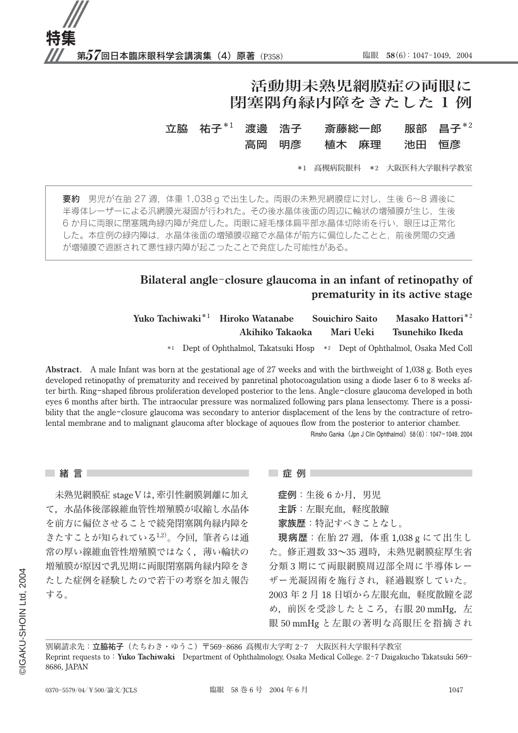
男児が在胎27週,体重1,038gで出生した。両眼の未熟児網膜症に対し,生後6~8週後に半導体レーザーによる汎網膜光凝固が行われた。その後水晶体後面の周辺に輪状の増殖膜が生じ,生後6か月に両眼に閉塞隅角緑内障が発症した。両眼に経毛様体扁平部水晶体切除術を行い,眼圧は正常化した。本症例の緑内障は,水晶体後面の増殖膜収縮で水晶体が前方に偏位したことと,前後房間の交通が増殖膜で遮断されて悪性緑内障が起こったことで発症した可能性がある。
A male Infant was born at the gestational age of 27 weeks and with the birthweight of 1,038g. Both eyes developed retinopathy of prematurity and received by panretinal photocoagulation using a diode laser 6 to 8 weeks after birth. Ring-shaped fibrous proliferation developed posterior to the lens. Angle-closure glaucoma developed in both eyes 6months after birth. The intraocular pressure was normalized following pars plana lensectomy. There is a possibility that the angle-closure glaucoma was secondary to anterior displacement of the lens by the contracture of retrolental membrane and to malignant glaucoma after blockage of aquoues flow from the posterior to anterior chamber.
Copyright © 2004, Igaku-Shoin Ltd. All rights reserved.