Japanese
English
- 有料閲覧
PPV購入案内
1,320円 (1,200円+税10%) こちらは、181ページから190ページの文献です。購入には医書.jp本会員登録が必要です。
会員登録完了後に改めて上記「購入サイトへ」を選択してください。
または「購入サイトへ」選択後に「購入ログイン」、「新規会員登録」の順に進んでください。
会員登録について詳しくはをご確認ください。
- Abstract 文献概要
- 1ページ目 Look Inside
- 参考文献 Reference
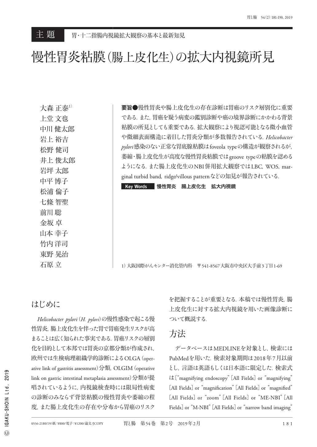
要旨●慢性胃炎や腸上皮化生の存在診断は胃癌のリスク層別化に重要である.また,胃癌を疑う病変の鑑別診断や癌の境界診断にかかわる背景粘膜の所見としても重要である.拡大観察により視認可能となる微小血管や微細表面構造に着目した胃炎分類が多数報告されている.Helicobacter pylori感染のない正常な胃底腺粘膜はfoveola typeの構造が観察されるが,萎縮・腸上皮化生が高度な慢性胃炎粘膜ではgroove typeの粘膜を認めるようになる.また腸上皮化生のNBI併用拡大観察ではLBC,WOS,marginal turbid band,ridge/villous patternなどの知見が報告されている.
It is essential to diagnose chronic gastritis and gastric intestinal metaplasia to stratify the risk of gastric cancer. Lesions representing chronic gastritis and gastric intestinal metaplasia require a differential diagnosis from gastric cancer. Gastric cancer is typically characterized by the presence of mucosa surrounding it, which is associated with its boundary diagnosis. Many studies have reported gastritis classifications on the basis of microvascular patterns and microsurface structures using magnifying endoscopy with NBI(narrow-band imaging). The normal gastric fundus gland mucosa without Helicobacter pylori infection has a foveola-type structure, whereas advanced chronic gastritis mucosa with severe atrophy and intestinal metaplasia has a groove-type structure. With the finding of intestinal metaplasia using magnifying endoscopy with NBI, several other findings such as a light-blue crest ; a white, opaque substance ; a marginal turbid band ; and a ridge/villous pattern have also been reported.
Copyright © 2019, Igaku-Shoin Ltd. All rights reserved.