Japanese
English
- 有料閲覧
- Abstract 文献概要
- 1ページ目 Look Inside
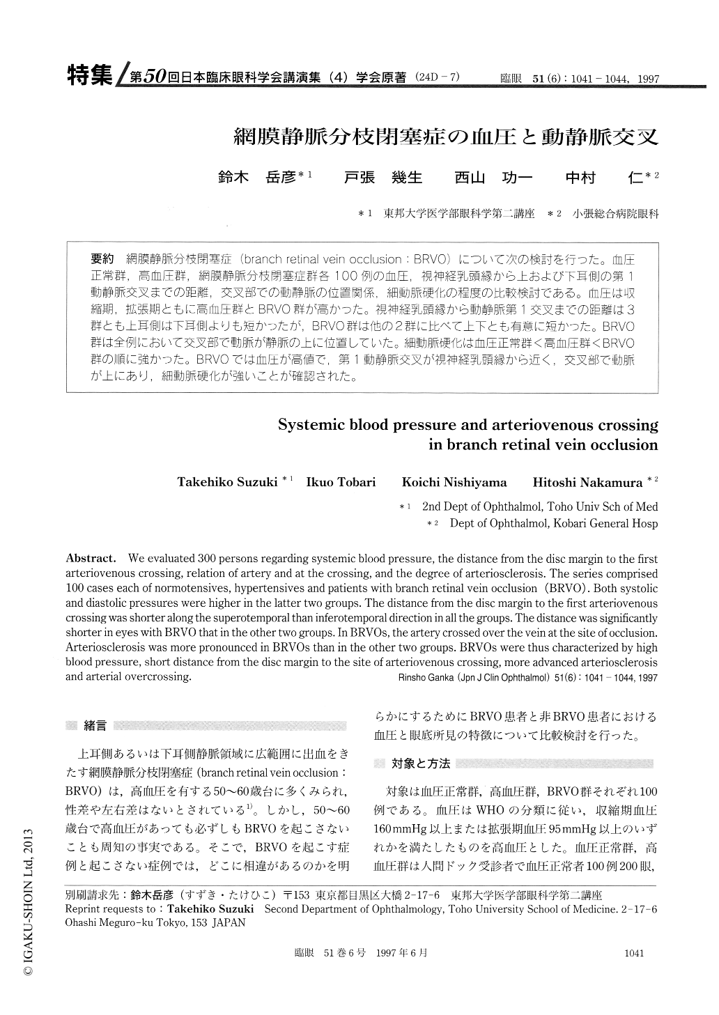
(24D-7) 網膜静脈分枝閉塞症(branch retina-vein occlusion:BRVO)について次の検討を行った。血圧正常群,高血圧群,網膜静脈分枝閉塞症群各100例の血圧,視神経乳頭縁から上および下耳側の第1動静脈交叉までの距離,交叉部での動静脈の位置関係,細動脈硬化の程度の比較検討である。血圧は収縮期,拡張期ともに高血圧群とBRVO群が高かった。視神経乳頭縁から動静脈第1交叉までの距離は3群とも上耳側は下耳側よりも短かったが,BRVO群は他の2群に比べて上下とも有意に短かった。BRVO群は全例において交叉部で動脈が静脈の上に位置していた。細動脈硬化は血圧正常群<高血圧群<BRVO群の順に強かった。BRVOでは血圧が高値で,第1動静脈交叉が視神経乳頭縁から近く,交叉部で動脈が上にあり,細動脈硬化が強いことが確認された。
We evaluated 300 persons regarding systemic blood pressure, the distance from the disc margin to the first arteriovenous crossing, relation of artery and at the crossing, and the degree of arteriosclerosis. The series comprised 100 cases each of normotensives, hypertensives and patients with branch retinal vein occlusion (BRVO). Both systolic and diastolic pressures were higher in the latter two groups. The distance from the disc margin to the first arteriovenous crossing was shorter along the superotemporal than inferotemporal direction in all the groups. The distance was significantly shorter in eyes with BRVO that in the other two groups. In BRVOs, the artery crossed over the vein at the site of occlusion. Arteriosclerosis was more pronounced in BRVOs than in the other two groups. BRVOs were thus characterized by high blood pressure, short distance from the disc margin to the site of arteriovenous crossing, more advanced arteriosclerosis and arterial overcrossing.

Copyright © 1997, Igaku-Shoin Ltd. All rights reserved.


