Japanese
English
- 有料閲覧
PPV購入案内
1,320円 (1,200円+税10%) こちらは、1079ページから1083ページの文献です。購入には医書.jp本会員登録が必要です。
会員登録完了後に改めて上記「購入サイトへ」を選択してください。
または「購入サイトへ」選択後に「購入ログイン」、「新規会員登録」の順に進んでください。
会員登録について詳しくはをご確認ください。
- Abstract 文献概要
- 1ページ目 Look Inside
I.はじめに
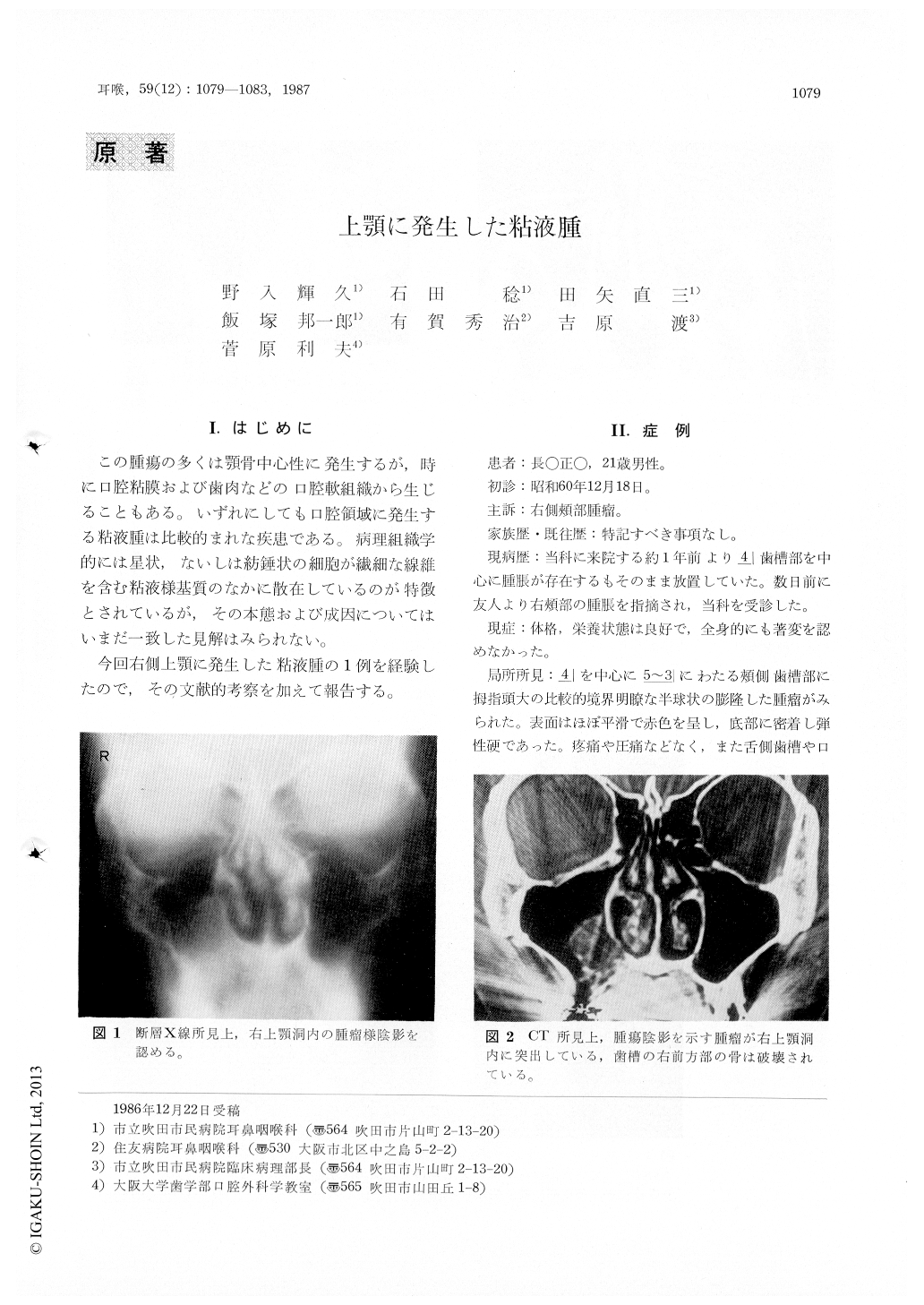
この腫瘍の多くは顎骨中心性に発生するが,時に口腔粘膜および歯肉などの口腔軟組織から生じることもある。いずれにしても口腔領域に発生する粘液腫は比較的まれな疾患である。病理組織学的には星状,ないしは紡錘状の細胞が繊細な線維を含む粘液様基質のなかに散在しているのが特徴とされているが,その本態および成因についてはいまだ一致した見解はみられない。
今回右側上顎に発生した粘液腫の1例を経験したので,その文献的考察を加えて報告する。
A 21-year-old male presented with a swelling without pain in the the right upper jaw. Radiographs demonstrated a large mass growing in the right maxillary sinus and a large radiolucent area of the right side of the upper jaw. The boundary of the lesion was comparatively clear radiographically. Removal of the tumor was performed under general anesthesia. The tumor was 4 × 4 × 2.5 cm in size, translucent and light brown in color, smooth surfaced with soft consistency and cystic lumens containing viscoid fluid. Microscopically, the tumor consisted of dense fibrous connective and myxomatous tissues. The tumor cells were stellate or spindle shaped, but mitosis was not seen. The histopathological diagnosis was myxoma. There has been no recurrence after treatment until present.
Copyright © 1987, Igaku-Shoin Ltd. All rights reserved.