Japanese
English
- 有料閲覧
PPV購入案内
1,320円 (1,200円+税10%) こちらは、563ページから566ページの文献です。購入には医書.jp本会員登録が必要です。
会員登録完了後に改めて上記「購入サイトへ」を選択してください。
または「購入サイトへ」選択後に「購入ログイン」、「新規会員登録」の順に進んでください。
会員登録について詳しくはをご確認ください。
- Abstract 文献概要
- 1ページ目 Look Inside
I.はじめに
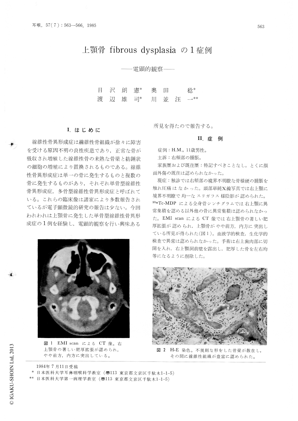
線維性骨異形成症は線維性骨組織が徐々に障害を受ける原因不明の良性疾患であり,正常な骨が吸収され増殖した線維性骨の未熟な骨梁と紡錘状の細胞の増殖により置換されるものである。線維性骨異形成症は単一の骨に発生するものと複数の骨に発生するものがあり,それぞれ単骨型線維性骨異形成症,多骨型線維性骨異形成症と呼ばれている。これらの臨床像は諸家により多数報告されているが電子顕微鏡的研究の報告は少ない。今回われわれは上顎骨に発生した単骨型線維性骨異形成症の1例を経験し,電顕的観察を行い興味ある所見を得たので報告する。
In a case of fibrous dysplasia of the maxillary bone, spindle cells were markedly increased in histological sections. The findings were characterized by oval irregular nucleus, actin-like filament, irregular basement lamina, and abandant and rough endoplasmic reticulum, ribosome, and glycogen granules. These cells seemed to be myofibroblasts, and microfibrils in the cytoplasma were numerous and released to the extracellular spaces, and then entered among collagen fibers. The arrangement of the collagen fibers was irregular and differed from that of normal fibrogenesis.
The presence of myofibroblasts and the abnormal fibrogenesis suggested to be one of the causes of fibrous dysplasia.
Copyright © 1985, Igaku-Shoin Ltd. All rights reserved.