Japanese
English
- 有料閲覧
PPV購入案内
1,320円 (1,200円+税10%) こちらは、165ページから173ページの文献です。購入には医書.jp本会員登録が必要です。
会員登録完了後に改めて上記「購入サイトへ」を選択してください。
または「購入サイトへ」選択後に「購入ログイン」、「新規会員登録」の順に進んでください。
会員登録について詳しくはをご確認ください。
- Abstract 文献概要
- 1ページ目 Look Inside
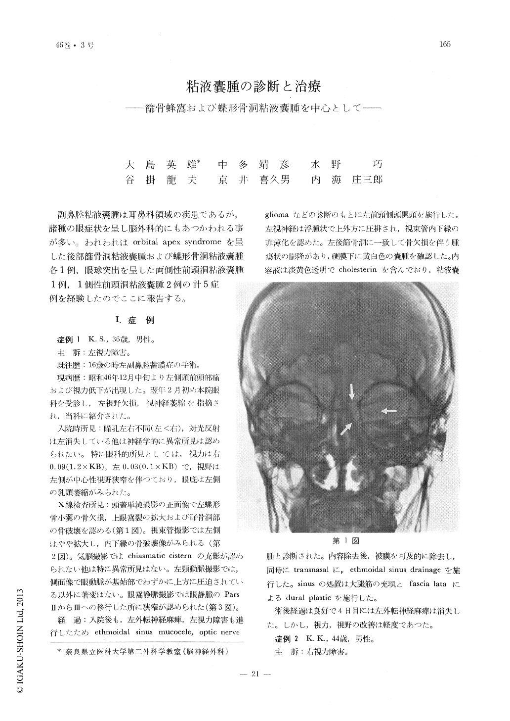
副鼻腔粘液嚢腫は耳鼻科領域の疾患であるが,諸種の眼症状を呈し脳外科的にもあつかわれる事が多い。われわれはorbital apex syndromeを呈した後部篩骨洞粘液嚢腫および蝶形骨洞粘液嚢腫各1例,眼球突出を呈した両側性前頭洞粘液嚢腫1例,1側性前頭洞粘液嚢腫2例の計5症例を経験したのでここに報告する。
Five cases of mucoceles, found in the following area, one each in the ethmoidal sinus, sphenoidal sinus, frontal sinus and both frontal sinuses in one case, are reported.
The mucocele of the posterior nasal sinuses is apt to be accompanied by orbital apex syndrome. Therefore it behooves the necessity of differenciating this affection with that of optic glioma, sphenoidal ridge meningioma, pituitary tumor, etc..
On the other hand, the mucocele of the frontal sinus may be easily diagnosed because of the fact it is always accompanied by symptoms of exophthalmos.
Most of the patients who suffer sinus mucocele complain of periorbital pain or headache.
A routine roentgenogram and tomogram will invariably reveal the evidence of enlargement or destruction of the bony structures of the sinus walls.
The surgical treatment of the mucocele is performed through transnasal or transcranial approach. In either case, the authors emphasize the necessity of establishing a good nasal drainage.
For the diagnosis and prognosis of the case the aid of the ophthalmologist may be required. The treatment should be established without delay.
Copyright © 1974, Igaku-Shoin Ltd. All rights reserved.