Japanese
English
- 有料閲覧
- Abstract 文献概要
- 1ページ目 Look Inside
Ⅰ.はじめに
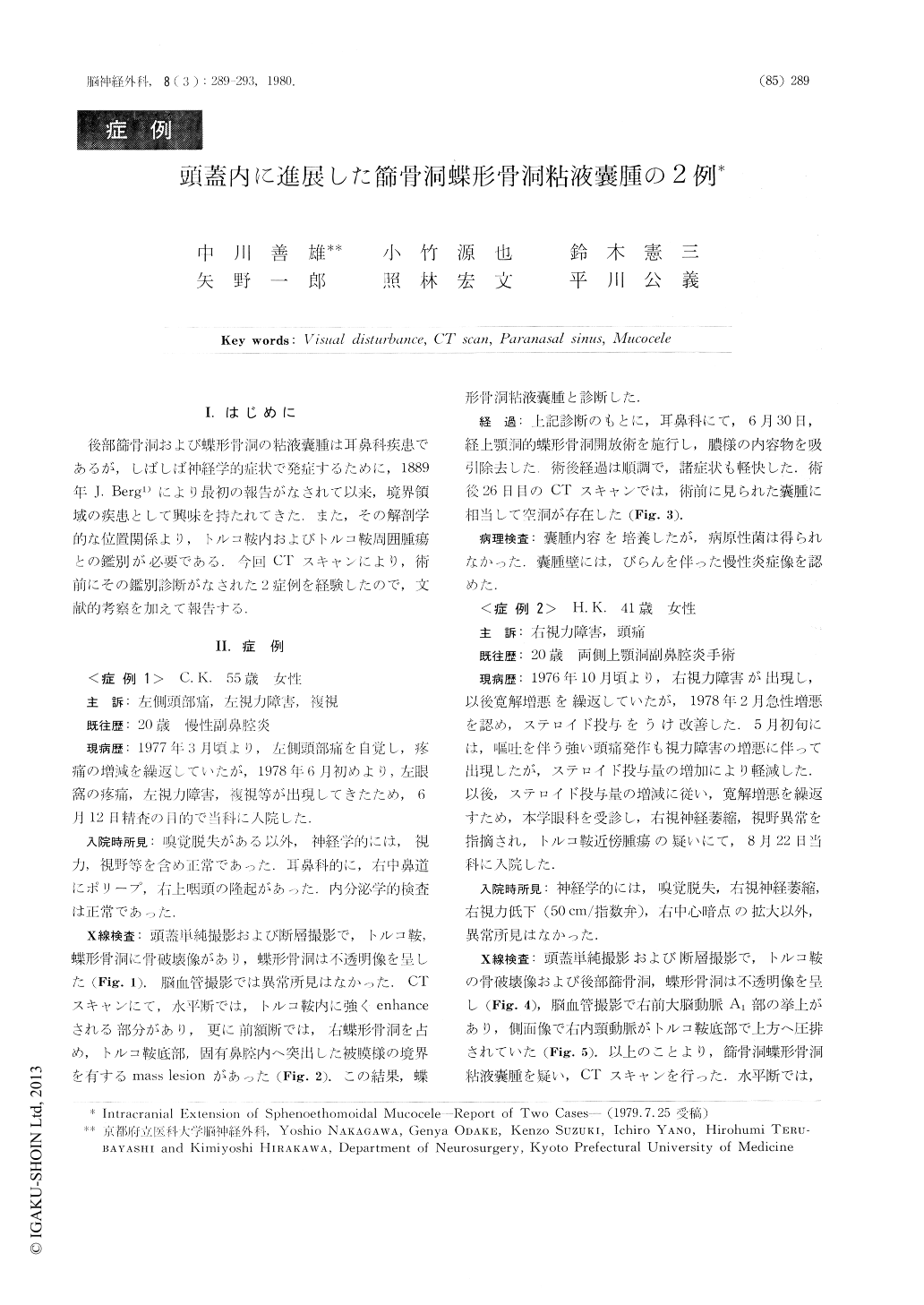
後部篩骨洞および蝶形骨洞の粘液嚢腫は耳鼻科疾患であるが,しばしば神経学的症状で発症するために,1889年J.Berg1)により最初の報告がなされて以来,境界領域の疾患として興味を持たれてきた.また,その解剖学的な位置関係より,トルコ鞍内およびトルコ鞍周囲腫瘍との鑑別が必要である.今回CTスキャンにより,術前にその鑑別診断がなされた2症例を経験したので,文献的考察を加えて報告する.
Two cases of sphenoethomoidal mucocele were report-ed. Both cases had had intermittent headaches and visual disturbances for few years. Its diagnosis was not clearly made by usual radiological examinations. But CT scans revealed intracranial mass lesion of homogeneous isoden-sity, with and without contrast medium injection (Houns-field Unit: 42), which extended from the sphenoid and ethomoid sinuses. This CT finding confirmed the diag-nosis of sphenoethomoidai mucocele.
The first case was a 55-year-old female. Her neuro-logicam examination revealed hyposmia.

Copyright © 1980, Igaku-Shoin Ltd. All rights reserved.


