Japanese
English
- 有料閲覧
PPV購入案内
1,320円 (1,200円+税10%) こちらは、27ページから34ページの文献です。購入には医書.jp本会員登録が必要です。
会員登録完了後に改めて上記「購入サイトへ」を選択してください。
または「購入サイトへ」選択後に「購入ログイン」、「新規会員登録」の順に進んでください。
会員登録について詳しくはをご確認ください。
- Abstract 文献概要
- 1ページ目 Look Inside
I.はじめに
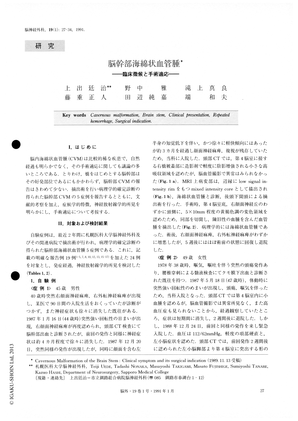
脳内海綿状血管腫(CVM)は比較的稀な疾患で,自然経過も明らかでなく,その手術適応に関しても議論の多いところである.とりわけ,橋をはじめとする脳幹部はその好発部位であるにもかかわらず,脳幹部CVMの報告はきわめて少ない.摘出術を行い病理学的確定診断の得られた脳幹部CVMの5症例を報告するとともに,文献的考察を加え,症候学的特徴,神経放射線学的所見を明らかにし,手術適応について考按する.
Clinical features and surgical indication of cavernous malformation (CVM) of the brain stem are reviewed, based on the analysis of five personally encountered cases and nineteen previously reported cases.
Nineteen out of these twenty four cases presented sudden onset of neurological deficits suggesting brain stem hemorrhage. Although the majority of those pa-tients with acute onset improved gradually within a couple of weeks, all cases, except for six cases treated with radical removal following their first bleeding epi-sodes, presented recurrent hemorrhages, and one third of them died due to the repeated hemorrhages. The other six cases presented progressive neurologic-al deficits with insidious onset. They were first dia-gnosed as brain stem gliomas or multiple sclerosis. All except two cases treated with radical removal of the CVM died within a year.
Surgical indication of the cerebral CVM is still con-troversial, because its clinical presentations and natural history have been unclear. Recent clinical application of magnetic resonance imaging enables us not only to make a preoperative diagnosis accurately, but also to evaluate the operability in relation to its anatomical location. Because of an apparent disposition towards re-peated hemorrhages, surgical removal should be consi-dered for the symptomatic CVM in the brain stem, on which microsurgical manipulation would be possible with an acceptable risk in many cases.
Copyright © 1991, Igaku-Shoin Ltd. All rights reserved.