Japanese
English
- 有料閲覧
PPV購入案内
1,320円 (1,200円+税10%) こちらは、1093ページから1098ページの文献です。購入には医書.jp本会員登録が必要です。
会員登録完了後に改めて上記「購入サイトへ」を選択してください。
または「購入サイトへ」選択後に「購入ログイン」、「新規会員登録」の順に進んでください。
会員登録について詳しくはをご確認ください。
- Abstract 文献概要
- 1ページ目 Look Inside
I.はじめに
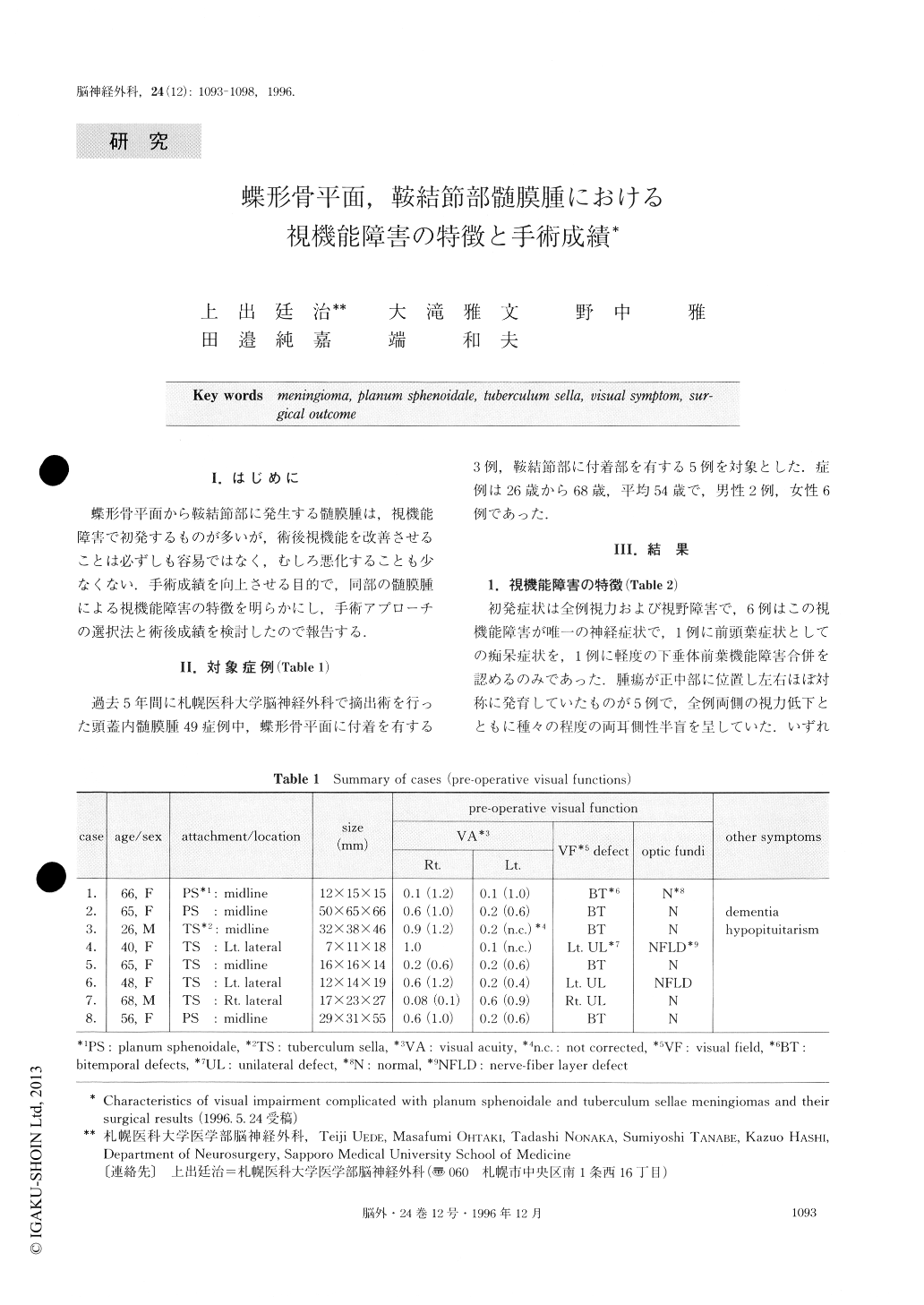
蝶形骨平面から鞍結節部に発生する髄膜腫は,視機能障害で初発するものが多いが,術後視機能を改善させることは必ずしも容易ではなく,むしろ悪化することも少なくない.手術成績を向上させる目的で,同部の髄膜腫による視機能障害の特徴を明らかにし,手術アプローチの選択法と術後成績を検討したので報告する.
It is well known that it is difficult to remove the pla-num sphenoidale and tuberculum sellae meningioma without damaging the optic nerves. The visual outcome after this operation has been unacceptable in such tumors, especially in large ones. This review propounds a strategy to secure visual acuity through operation.
A total of eight cases are summarized. In five midline symmetrical meningiomas, the tumors compressed the nerves at the portion of the optic chiasma, causing a typical bitemporal hemianopsia. Four large tumors were resected by the frontobasal interhemispheric approach to minimize the intraoperative damage to the optic chiasma, and a small one was removed by the pterional approach. Visual deficits were recovered immediately after the operation in all cases without any surgical complications.
Three meningiomas were attached to the lateral part of the planum sphenoidale or tuberculum sella. Although the sizes were relatively small in all cases, they caused ipsilateral severe visual loss by direct com-pression to optic nerves. MRI and three-dimensional CT angiography showed the tumor extension into the optic canal. The ipsilateral pterional approach was selected in these cases. To avoid additional nerve dam-age, we tried to reduce the tension of nerves which were compressed by the tumors. We removed the ante-rior clinoid process and opened the optic canal before surgical manipulation of the tumor. In two cases, tumors severely compressed the optic nerves from the medial side, and nerves were stretched laterally. Great care was required to separate the optic nerves from tumors in those two cases. In contrast, the resection seemed to be very easy in one of the cases where the optic nerve was displaced infero-medially. Visual symp-toms were improved in all cases, although one case be-came worse temporarily.
Although planum sphenoidale and tuberculum sellae meningiomas are still troublesome, appropriate preop-erative management would allow us to expect an excel-lent visual outcome. Especially, selection of the surgical approach should be based on the anatomical analysis of the nerve displacement.
Copyright © 1996, Igaku-Shoin Ltd. All rights reserved.