Japanese
English
- 有料閲覧
PPV購入案内
1,320円 (1,200円+税10%) こちらは、83ページから87ページの文献です。購入には医書.jp本会員登録が必要です。
会員登録完了後に改めて上記「購入サイトへ」を選択してください。
または「購入サイトへ」選択後に「購入ログイン」、「新規会員登録」の順に進んでください。
会員登録について詳しくはをご確認ください。
- Abstract 文献概要
- 1ページ目 Look Inside
I.はじめに
海綿状血管腫は中枢神経系のvascular malformationの5-13%を占めると言われており18),比較的稀な疾患とされている.特に脳幹部に存在するものは稀で,かつては生前診断は困難であった.
しかし,MRI導入後その特徴ある所見より比較的容易に診断されるようになり,出血を繰り返す例ではその局在により外科的切除が行われる報告が増加している3,5,8-13,19-23).
今回われわれは手術的に全摘し得た橋外側部の海綿状血管腫を経験したので,今までに報告されている脳幹部海綿状血管腫の手術例を含わせ,外科的治療の成績について報告する.
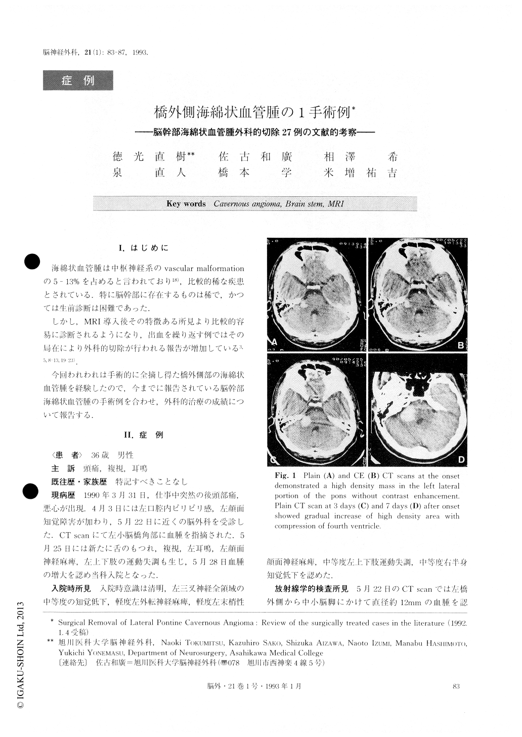
A case of cavernous angioma of the pons which was surgically and successfully excised was reported. A 36 year-old man complained of progressive
headache, double vision and tinnitis. Neurologic ex-amination revealed left fifth, sixth and seventh cranial nerve palsies. He had left limb ataxia and right sided hemisensory deficit. A computed tomographic (CT) scan on admission disclosed a hematoma in the left lateral portion of the puns. Serial CT scans demonstrated pro-gressive increase of hematoma. MRI scans revealed an area of mixed signal intensity in TI weighted images. These findings were thought to be consistent with a cavernous angioma. Three months after the onset, surgery was performed using a lateral suboccipital approach. Histological examination disclosed cavernous angioma. After surgery, the patient's neurological de-ficits improved, and after 3 months, all symptoms except the mild limb ataxia had disappeared.
Copyright © 1993, Igaku-Shoin Ltd. All rights reserved.