Japanese
English
- 有料閲覧
PPV購入案内
1,320円 (1,200円+税10%) こちらは、640ページから650ページの文献です。購入には医書.jp本会員登録が必要です。
会員登録完了後に改めて上記「購入サイトへ」を選択してください。
または「購入サイトへ」選択後に「購入ログイン」、「新規会員登録」の順に進んでください。
会員登録について詳しくはをご確認ください。
- Abstract 文献概要
- 1ページ目 Look Inside
Ⅰ.序
Wilson病脳に出現するOpalski細胞は,その特異な大きさと細胞質の性状から,Opalski1)の報告以来多くの関心が寄せられ,その起原と木態,AlzheimerⅠ型グリアとの関係,さらにWilson病における特異性などについて多数の研究がなされてきたが,いまだにこれらの点について意見の一致は認められない。
これらの従来の研究は,すべてNissl染色や鍍銀法標本などの光学顕微鏡によるものであり,Opalski細胞に関する電顕的観察は著者らによる報告2)があるのみである。
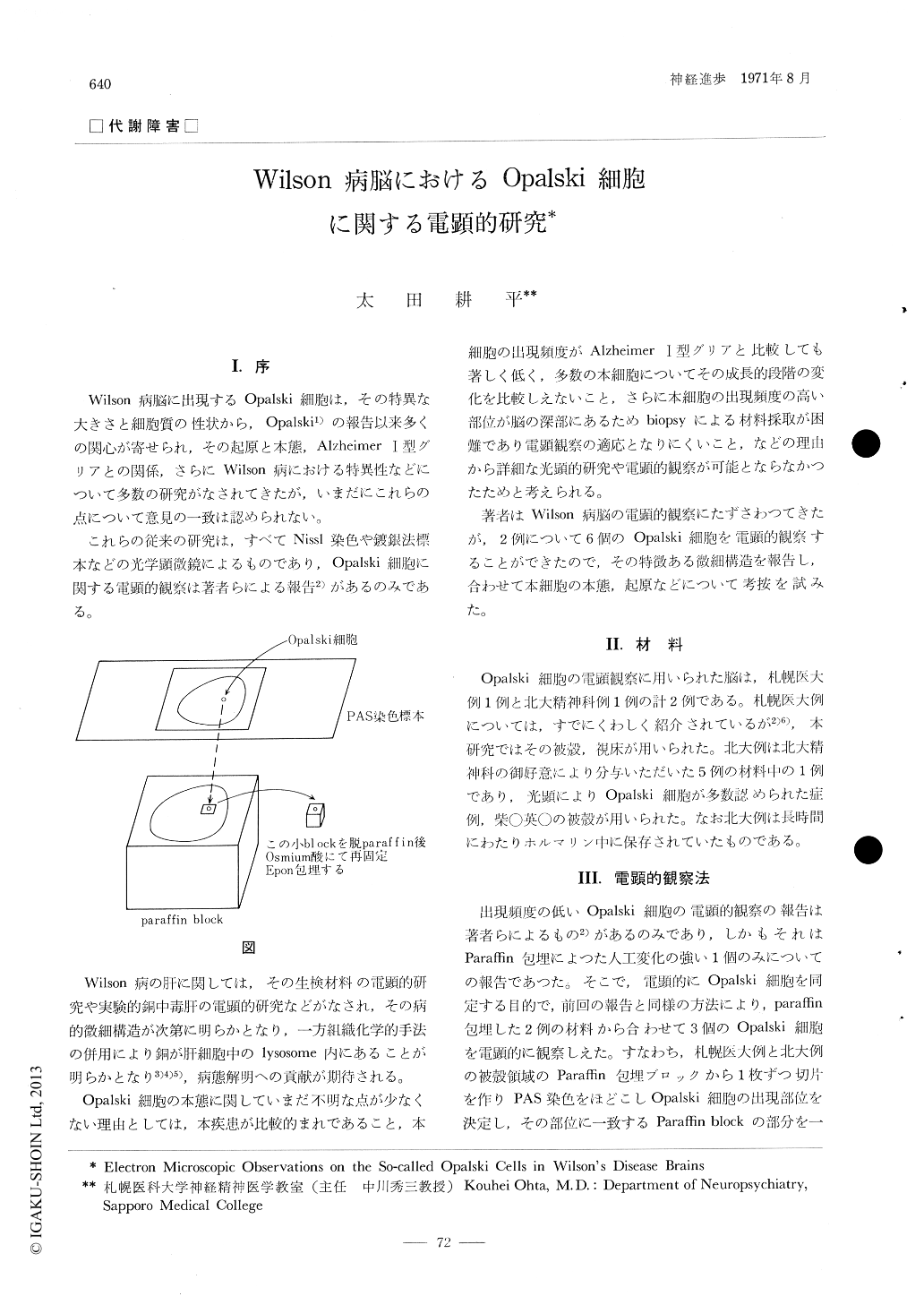
The ultrastructures of Opalski cells in two cases of Wilson's disease were observed by electron microscope. Identification of Opalski cells was performed under specical care using paraffin embedded materials. The surface area of the paraffin block on which the Opalski cell existed was determined with PAS-stained sections light microscopically, then the area containing the Opalski cell was cut off as a small square paraffin block. After the small square block was deparaffinized, it was refixed with osmium tetroxide and embedded in Epon.
Copyright © 1971, Igaku-Shoin Ltd. All rights reserved.