Japanese
English
- 有料閲覧
PPV購入案内
1,320円 (1,200円+税10%) こちらは、665ページから676ページの文献です。購入には医書.jp本会員登録が必要です。
会員登録完了後に改めて上記「購入サイトへ」を選択してください。
または「購入サイトへ」選択後に「購入ログイン」、「新規会員登録」の順に進んでください。
会員登録について詳しくはをご確認ください。
- Abstract 文献概要
- 1ページ目 Look Inside
はじめに
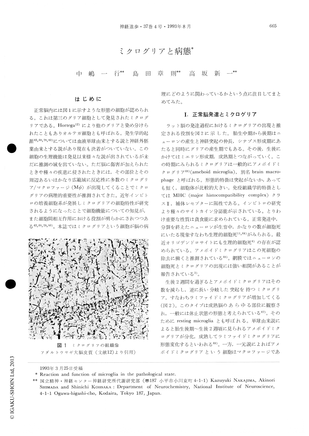
正常脳内には図1に示すような形態の細胞が認められる。これは第三のグリア細胞として発見されたミクログリアである。Hortega12)により他のグリアと染め分けられたこともありオルテガ細胞とも呼ばれる。発生学的起源42,68,78,80)については血液単球由来とする説と神経外胚葉由来とする説があり現在も決着がついていない。この細胞の生理機能は発見以来様々な説が出されているが未だに推測の域を出ていない。ただ脳に傷害が加えられたときや種々の疾患に侵されたときには,その部位とその周辺あるいはかなり広範域に反応性に多数のミクログリア/マクロファージ(Mφ)が出現してくることでミクログリアの病理的重要性が推測されてきた。近年インビトロの培養細胞系が発展しミクログリアの細胞特性が研究されるようになったことで細胞機能についての知見が,また細胞間相互作用における役割が明らかにされつつある42,68,78,80)。本誌ではミクログリアという細胞が脳の病理にどのように関わっているかという点に注目してまとめてみた。
Microglia, a type of glial cells in the central nervous system (CNS), are distributed as ramified microglia with a certain regular spaces in normal adult brain parenchyma, and is generally believed to be transformed from ameboid microglia (brain macrophages). But, their ontogenic origin is now controversial; there are two lines, monocyte-methodermal and neuroectodermal origin.
Once brain is injured or taken brain disease, microglia are activated, proliferated and accumulated in the injured sites. These reactive microglia are thought to be involved in defense against the infec-tion, phagocytic scavenge of dead cells, antigen presentation, gliosis and tissue repair. However, CNS injures and most of brain diseases are usually accompanied by disruption of the vasculature and a following invasion of blood-derived monocytes and macrophages into the parenchyma. Since there is no microglia specific markers which can distinguish from other peripheral macrophages, it is difficult to observe only microglial reactions or roles in injured sites.
Copyright © 1993, Igaku-Shoin Ltd. All rights reserved.