Japanese
English
- 有料閲覧
PPV購入案内
1,320円 (1,200円+税10%) こちらは、377ページから380ページの文献です。購入には医書.jp本会員登録が必要です。
会員登録完了後に改めて上記「購入サイトへ」を選択してください。
または「購入サイトへ」選択後に「購入ログイン」、「新規会員登録」の順に進んでください。
会員登録について詳しくはをご確認ください。
- Abstract 文献概要
- 1ページ目 Look Inside
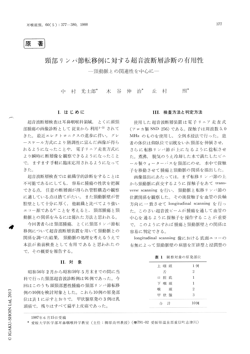
I.はじめに
超音波断層検査は耳鼻咽喉科領域,とくに頭頸部腫瘍の画像診断として従来から利用1〜3)されてきた。最近エレクトロニクスの進歩に伴い,グレースケール方式により階調性に富んだ画像が得られるようになったことや,電子リニア走査方式により瞬時に断層像を観察できるようになったことで,ますます手軽に臨床応用されるようになってきた。
超音波断層検査では組織学的診断をすることは不可能であるにしても,容易に腫瘍の性状を把握できる点,任意の断層面が得られ管腔構造の観察に適している点は捨てがたい。また頸動脈壁が管腔壁として十分に厚く,他組織と比べてより強いエコー源である4)ことを考えると,頸部腫瘍と頸動脈との関係をみるには優れた方法と思われる。
Using an electronic linear scanning ultrasonic echography, the relationship between metastatic lymph nodes and the carotid artery was observed in ten patients. The longitudinal scanning was utilized along the carotid artery. According to the echo pattern from tumor base, the echographic findings were classified into a compression type and an infiltration type. The former type was seen in seven patients and the latter in one, whose carotid artery was surgically confirmed to be in-filtrated by the tumor. As the preoperative in-vestigation of the wall of the carotid artery, this ultrasonic echography was more advantageous than other examinations.
Copyright © 1988, Igaku-Shoin Ltd. All rights reserved.