Japanese
English
- 有料閲覧
PPV購入案内
1,320円 (1,200円+税10%) こちらは、817ページから821ページの文献です。購入には医書.jp本会員登録が必要です。
会員登録完了後に改めて上記「購入サイトへ」を選択してください。
または「購入サイトへ」選択後に「購入ログイン」、「新規会員登録」の順に進んでください。
会員登録について詳しくはをご確認ください。
- Abstract 文献概要
- 1ページ目 Look Inside
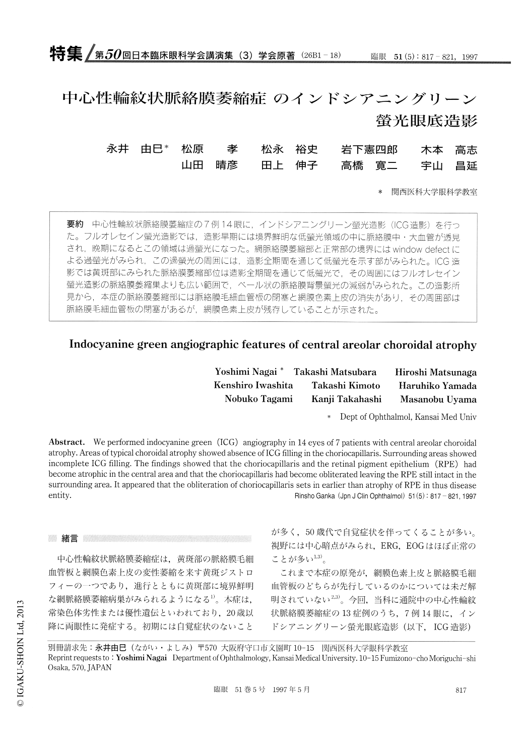
(26B1-18) 中心性輪紋状脈絡膜萎縮症の7例14眼に,インドシアニングリーン螢光造影(ICG造影)を行った。フルオレセイン螢光造影では,造影早期には境界鮮明な低螢光領域の中に脈絡膜中・大血管が透見され,晩期になるとこの領域は過螢光になった。網脈絡膜萎縮部と正常部の境界にはwindow defectによる過螢光がみられ,この過螢光の周囲には,造影全期間を通じて低螢光を示す部がみられた。ICG造影では黄斑部にみられた脈絡膜萎縮部位は造影全期間を通じて低螢光で,その周囲にはフルオレセイン螢光造影の脈絡膜萎縮巣よりも広い範囲で,ベール状の脈絡膜背景螢光の減弱がみられた。この造影所見から.本症の脈絡膜萎縮部には脈絡膜毛細血管板の閉塞と網膜色素上皮の消失があり,その周囲部は脈絡膜毛細血管板の閉塞があるが,網膜色素上皮が残存していることが示された。
We performed indocyanine green (ICG) angiography in 14 eyes of 7 patients with central areolar choroidal atrophy. Areas of typical choroidal atrophy showed absence of ICG filling in the choriocapillaris. Surrounding areas showed incomplete ICG filling. The findings showed that the choriocapillaris and the retinal pigment epithelium (RPE) had become atrophic in the central area and that the choriocapillaris had become obliterated leaving the RPE still intact in the surrounding area. It appeared that the obliteration of choriocapillaris sets in earlier than atrophy of RPE in thus disease entity.
Copyright © 1997, Igaku-Shoin Ltd. All rights reserved.