Japanese
English
- 有料閲覧
- Abstract 文献概要
- 1ページ目 Look Inside
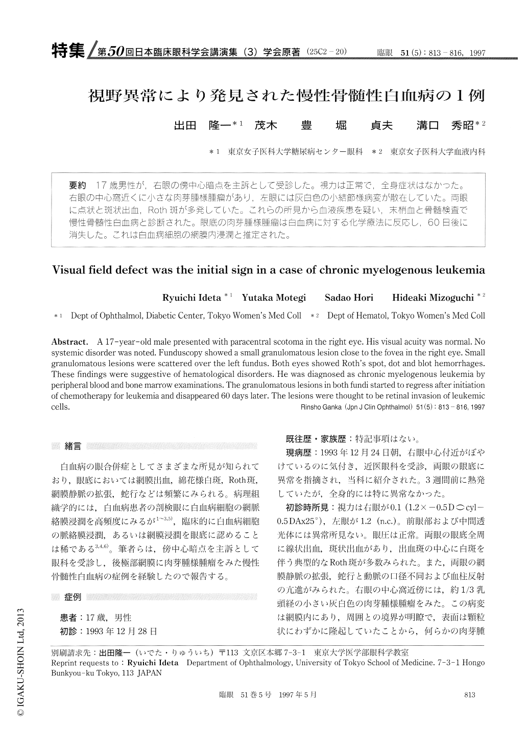
(25C-20) 17歳男性が,右眼の傍中心暗点を主訴として受診した。視力は正常で,全身症状はなかった。右眼の中心窩近くに小さな肉芽腫様腫瘤があり.左眼には灰白色の小結節様病変が散在していた。両眼に点状と斑状出血,Roth斑が多発していた。これらの所見から血液疾患を疑い,末梢血と骨髄検査で慢性骨髄性白血病と診断された。眼底の肉芽腫様腫瘤は白血病に対する化学療法に反応し,60日後に消失した。これは白血病細胞の網膜内浸潤と推定された。
A 17-year-old male presented with paracentral scotoma in the right eye. His visual acuity was normal. No systemic disorder was noted. Funduscopy showed a small granulomatous lesion close to the fovea in the right eye. Small granulomatous lesions were scattered over the left fundus. Both eyes showed Roth's spot, dot and blot hemorrhages. These findings were suggestive of hematological disorders. He was diagnosed as chronic myelogenous leukemia by peripheral blood and bone marrow examinations. The granulomatous lesions in both fundi started to regress after initiation of chemotherapy for leukemia and disappeared 60 days later. The lesions were thought to be retinal invasion of leukemic cells.

Copyright © 1997, Igaku-Shoin Ltd. All rights reserved.


