Japanese
English
- 有料閲覧
PPV購入案内
1,320円 (1,200円+税10%) こちらは、569ページから574ページの文献です。購入には医書.jp本会員登録が必要です。
会員登録完了後に改めて上記「購入サイトへ」を選択してください。
または「購入サイトへ」選択後に「購入ログイン」、「新規会員登録」の順に進んでください。
会員登録について詳しくはをご確認ください。
- Abstract 文献概要
- 1ページ目 Look Inside
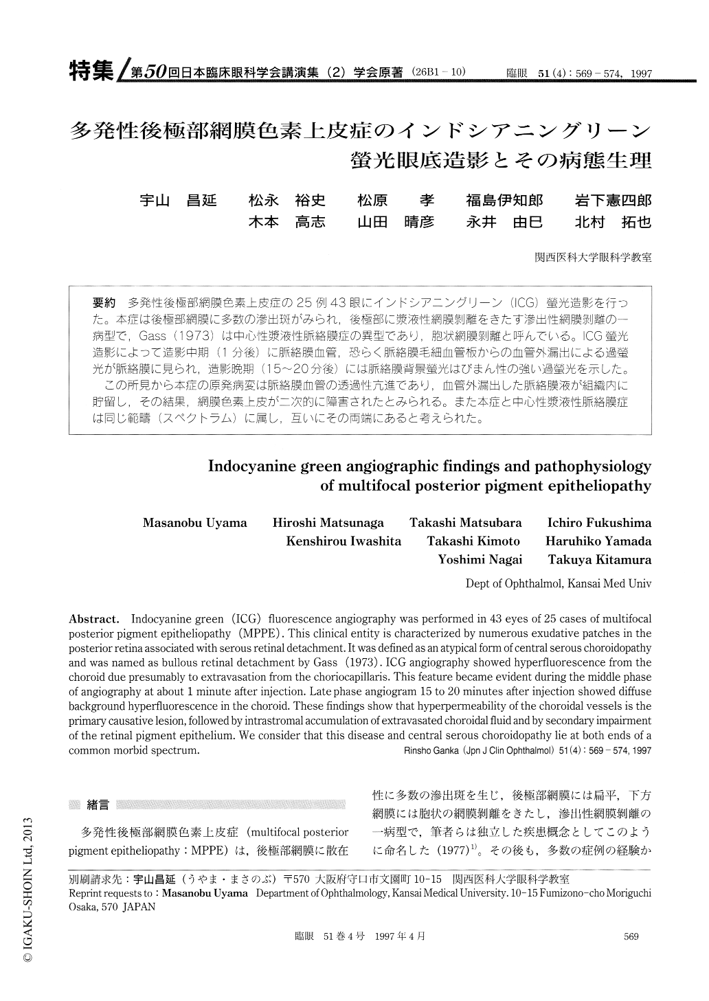
(26B1-10) 多発性後極部網膜色素上皮症の25例43眼にインドシアニングリーン(lCG)螢光造影を行った。本症は後極部網膜に多数の滲出斑がみられ,後極部に漿液性網膜剥離をきたす滲出性網膜剥離の一病型で,Gass (1973)は中心性漿液性脈絡膜症の異型であり,胞状網膜剥離と呼んでいる。ICG螢光造影によって造影中期(1分後)に脈絡膜血管,恐らく脈絡膜毛細血管板からの血管外漏出による過螢光が脈絡膜に見られ,造影晩期(15〜20分後)には脈絡膜背景螢光はびまん性の強い過螢光を示した。
この所見から本症の原発病変は脈絡膜血管の透過性亢進であり,血管外漏出した脈絡膜液が組織内に貯留し,その結果,網膜色素上皮が二次的に障害されたとみられる。また本症と中心性漿液性脈絡膜症は同じ範疇(スペクトラム)に属し,互いにその両端にあると考えられた。
Indocyanine green (ICG) fluorescence angiography was performed in 43 eyes of 25 cases of multifocal posterior pigment epitheliopathy (MPPE) . This clinical entity is characterized by numerous exudative patches in the posterior retina associated with serous retinal detachment. It was defined as an atypical form of central serous choroidopathy and was named as bullous retinal detachment by Gass (1973) . ICG angiography showed hyperfluorescence from the choroid due presumably to extravasation from the choriocapillaris. This feature became evident during the middle phase of angiography at about 1 minute after injection. Late phase angiogram 15 to 20 minutes after injection showed diffuse background hyperfluorescence in the choroid. These findings show that hyperpermeability of the choroidal vessels is the primary causative lesion, followed by intrastromal accumulation of extravasated choroidal fluid and by secondary impairment of the retinal pigment epithelium. We consider that this disease and central serous choroidopathy lie at both ends of a common morbid spectrum.
Copyright © 1997, Igaku-Shoin Ltd. All rights reserved.