Japanese
English
- 有料閲覧
PPV購入案内
1,320円 (1,200円+税10%) こちらは、1094ページから1098ページの文献です。購入には医書.jp本会員登録が必要です。
会員登録完了後に改めて上記「購入サイトへ」を選択してください。
または「購入サイトへ」選択後に「購入ログイン」、「新規会員登録」の順に進んでください。
会員登録について詳しくはをご確認ください。
- Abstract 文献概要
- 1ページ目 Look Inside
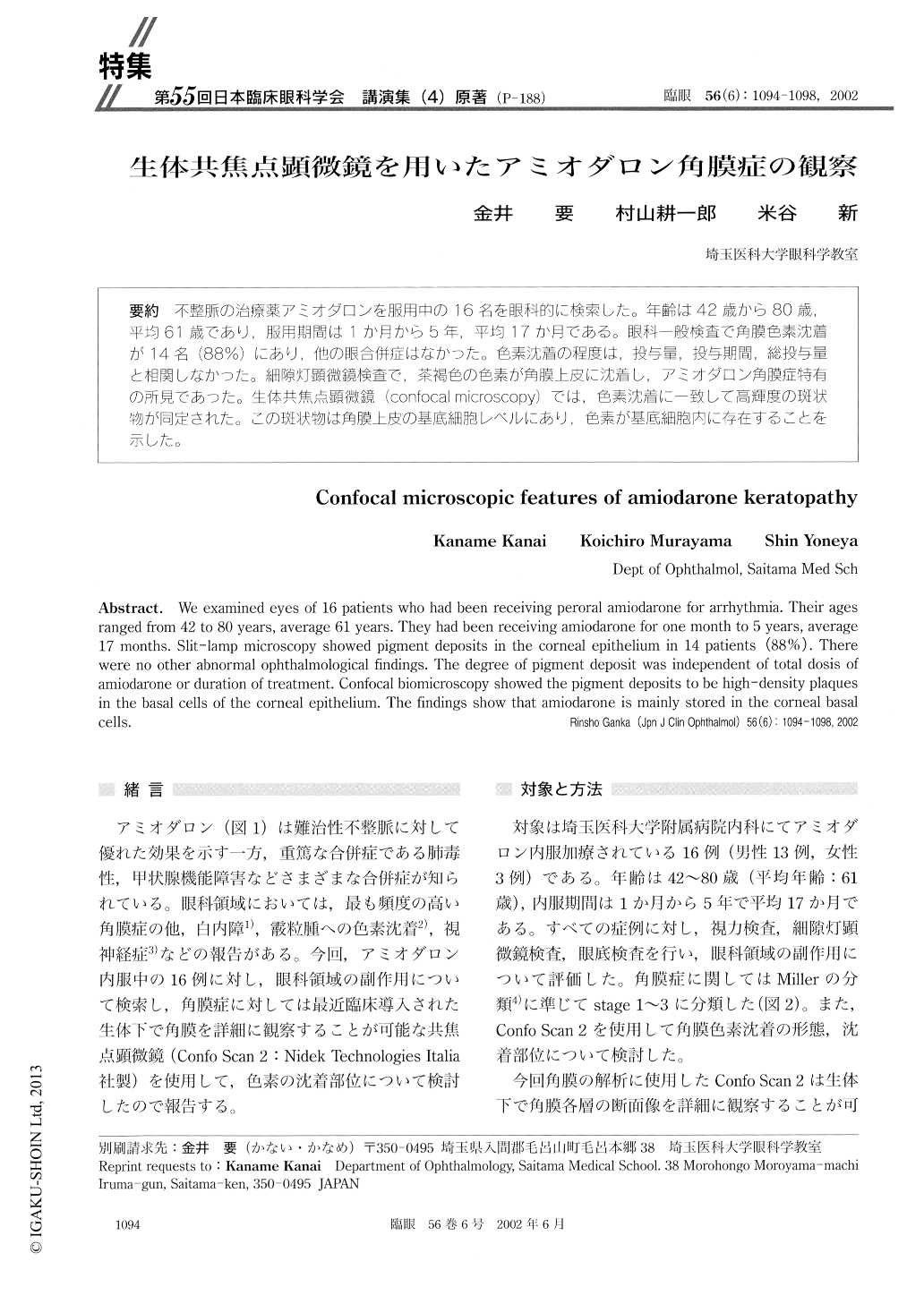
不整脈の治療薬アミオダロンを服用中の16名を眼科的に検索した。年齢は42歳から80歳,平均61歳であり,服用期間は1か月から5年,平均17か月である。眼科一般検査で角膜色素沈着が14名(88%)にあり,他の眼合併症はなかった。色素沈着の程度は,投与量,投与期間,総投与量と相関しなかった。細隙灯顕微鏡検査で,茶褐色の色素が角膜上皮に沈着し,アミオダロン角膜症特有の所見であった。生体共焦点顕微鏡(confocal microscopy)では,色素沈着に一致して高輝度の斑状物が同定された。この斑状物は角膜上皮の基底細胞レベルにあり,色素が基底細胞内に存在することを示した。
We examined eyes of 16 patients who had been receiving peroral amiodarone for arrhythmia. Their ages ranged from 42 to 80 years, average 61 years. They had been receiving amiodarone for one month to 5 years, average 17 months. Slit-lamp microscopy showed pigment deposits in the corneal epithelium in 14 patients (88%). There were no other abnormal ophthalmological findings. The degree of pigment deposit was independent of total dosis of amiodarone or duration of treatment. Confocal biomicroscopy showed the pigment deposits to be high-density plaques in the basal cells of the corneal epithelium. The findings show that amiodarone is mainly stored in the corneal basal cells.
Copyright © 2002, Igaku-Shoin Ltd. All rights reserved.