Japanese
English
- 有料閲覧
PPV購入案内
1,320円 (1,200円+税10%) こちらは、983ページから988ページの文献です。購入には医書.jp本会員登録が必要です。
会員登録完了後に改めて上記「購入サイトへ」を選択してください。
または「購入サイトへ」選択後に「購入ログイン」、「新規会員登録」の順に進んでください。
会員登録について詳しくはをご確認ください。
- Abstract 文献概要
- 1ページ目 Look Inside
- 参考文献 Reference
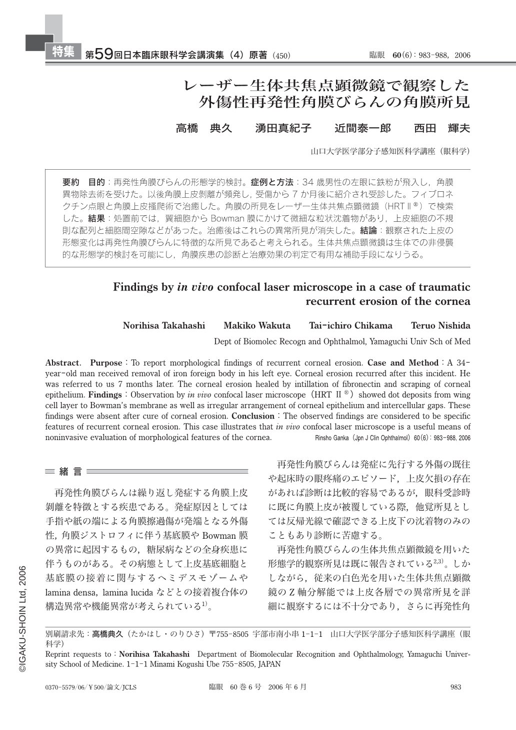
目的:再発性角膜びらんの形態学的検討。症例と方法:34歳男性の左眼に鉄粉が飛入し,角膜異物除去術を受けた。以後角膜上皮剝離が頻発し,受傷から7か月後に紹介され受診した。フィブロネクチン点眼と角膜上皮搔爬術で治癒した。角膜の所見をレーザー生体共焦点顕微鏡(HRTⅡ(R))で検索した。結果:処置前では,翼細胞からBowman膜にかけて微細な粒状沈着物があり,上皮細胞の不規則な配列と細胞間空隙などがあった。治癒後はこれらの異常所見が消失した。結論:観察された上皮の形態変化は再発性角膜びらんに特徴的な所見であると考えられる。生体共焦点顕微鏡は生体での非侵襲的な形態学的検討を可能にし,角膜疾患の診断と治療効果の判定で有用な補助手段になりうる。
Purpose:To report morphological findings of recurrent corneal erosion. Case and Method:A 34-year-old man received removal of iron foreign body in his left eye. Corneal erosion recurred after this incident. He was referred to us 7months later. The corneal erosion healed by intillation of fibronectin and scraping of corneal epithelium. Findings:Observation by in vivo confocal laser microscope(HRT Ⅱ(R))showed dot deposits from wing cell layer to Bowman's membrane as well as irregular arrangement of corneal epithelium and intercellular gaps. These findings were absent after cure of corneal erosion. Conclusion:The observed findings are considered to be specific features of recurrent corneal erosion. This case illustrates that in vivo confocal laser microscope is a useful means of noninvasive evaluation of morphological features of the cornea.
Copyright © 2006, Igaku-Shoin Ltd. All rights reserved.