Japanese
English
- 有料閲覧
PPV購入案内
1,320円 (1,200円+税10%) こちらは、915ページから920ページの文献です。購入には医書.jp本会員登録が必要です。
会員登録完了後に改めて上記「購入サイトへ」を選択してください。
または「購入サイトへ」選択後に「購入ログイン」、「新規会員登録」の順に進んでください。
会員登録について詳しくはをご確認ください。
- Abstract 文献概要
- 1ページ目 Look Inside
- 参考文献 Reference
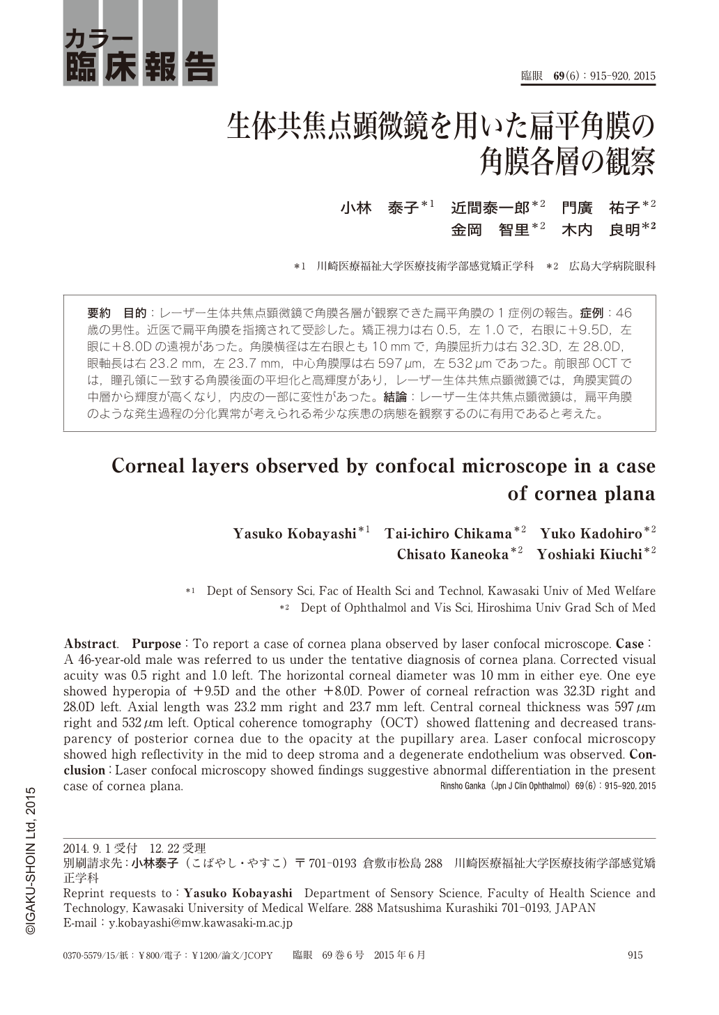
要約 目的:レーザー生体共焦点顕微鏡で角膜各層が観察できた扁平角膜の1症例の報告。症例:46歳の男性。近医で扁平角膜を指摘されて受診した。矯正視力は右0.5,左1.0で,右眼に+9.5D,左眼に+8.0Dの遠視があった。角膜横径は左右眼とも10mmで,角膜屈折力は右32.3D,左28.0D,眼軸長は右23.2mm,左23.7mm,中心角膜厚は右597μm,左532μmであった。前眼部OCTでは,瞳孔領に一致する角膜後面の平坦化と高輝度があり,レーザー生体共焦点顕微鏡では,角膜実質の中層から輝度が高くなり,内皮の一部に変性があった。結論:レーザー生体共焦点顕微鏡は,扁平角膜のような発生過程の分化異常が考えられる希少な疾患の病態を観察するのに有用であると考えた。
Abstract. Purpose:To report a case of cornea plana observed by laser confocal microscope. Case:A 46-year-old male was referred to us under the tentative diagnosis of cornea plana. Corrected visual acuity was 0.5 right and 1.0 left. The horizontal corneal diameter was 10 mm in either eye. One eye showed hyperopia of +9.5D and the other +8.0D. Power of corneal refraction was 32.3D right and 28.0D left. Axial length was 23.2 mm right and 23.7 mm left. Central corneal thickness was 597μm right and 532μm left. Optical coherence tomography(OCT)showed flattening and decreased transparency of posterior cornea due to the opacity at the pupillary area. Laser confocal microscopy showed high reflectivity in the mid to deep stroma and a degenerate endothelium was observed. Conclusion:Laser confocal microscopy showed findings suggestive abnormal differentiation in the present case of cornea plana.
Copyright © 2015, Igaku-Shoin Ltd. All rights reserved.