Japanese
English
- 有料閲覧
PPV購入案内
1,320円 (1,200円+税10%) こちらは、403ページから407ページの文献です。購入には医書.jp本会員登録が必要です。
会員登録完了後に改めて上記「購入サイトへ」を選択してください。
または「購入サイトへ」選択後に「購入ログイン」、「新規会員登録」の順に進んでください。
会員登録について詳しくはをご確認ください。
- Abstract 文献概要
- 1ページ目 Look Inside
- 参考文献 Reference
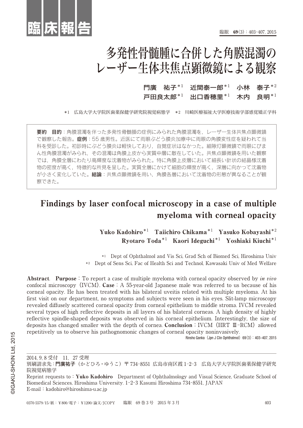
要約 目的:角膜混濁を伴った多発性骨髄腫の症例にみられた角膜混濁を,レーザー生体共焦点顕微鏡で観察した報告。症例:55歳男性。近医にて両眼ぶどう膜炎加療中に両眼の角膜変性症を疑われて当科を受診した。初診時にぶどう膜炎は軽快しており,自覚症状はなかった。細隙灯顕微鏡で両眼にびまん性角膜混濁がみられ,その混濁は角膜上皮から実質中層に散在していた。共焦点顕微鏡を用いた観察では,角膜全層にわたり高輝度な沈着物がみられた。特に角膜上皮層において細長い針状の結晶様沈着物の密度が高く,特徴的な所見を呈した。実質全層にかけて細胞の輝度が高く,深層に向かって沈着物が小さく変化していた。結論:共焦点顕微鏡を用い,角膜各層において沈着物の形態が異なることが観察できた。
Abstract. Purpose:To report a case of multiple myeloma with corneal opacity observed by in vivo confocal microscopy(IVCM). Case:A 55-year-old Japanese male was referred to us because of his corneal opacity. He has been treated with his bilateral uveitis related with multiple myeloma. At his first visit on our department, no symptoms and subjects were seen in his eyes. Slit-lamp microscopy revealed diffusely scattered corneal opacity from corneal epithelium to middle stroma. IVCM revealed several types of high reflective deposits in all layers of his bilateral corneas. A high density of highly reflective spindle-shaped deposits was observed in his corneal epithelium. Interestingly, the size of deposits has changed smaller with the depth of cornea. Conclusion:IVCM(HRT Ⅲ-RCM)allowed repetitively us to observe his pathognomonic changes of corneal opacity noninvasively.
Copyright © 2015, Igaku-Shoin Ltd. All rights reserved.