Japanese
English
- 有料閲覧
PPV購入案内
1,320円 (1,200円+税10%) こちらは、1847ページから1851ページの文献です。購入には医書.jp本会員登録が必要です。
会員登録完了後に改めて上記「購入サイトへ」を選択してください。
または「購入サイトへ」選択後に「購入ログイン」、「新規会員登録」の順に進んでください。
会員登録について詳しくはをご確認ください。
- Abstract 文献概要
- 1ページ目 Look Inside
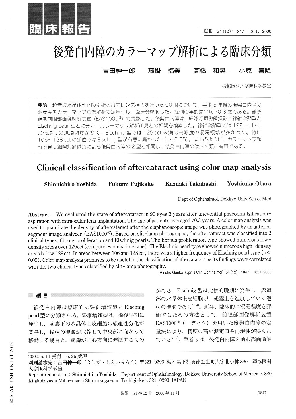
超音波水晶体乳化吸引術と眼内レンズ挿入を行った90眼について,手術3年後の後発白内障の混濁度をカラーマップ画像解析で定量化し,臨床分類をした。症例の年齢は平均70.3歳である。徹照像を前眼部画像解析装置(EAS1000®)で撮影した。後発白内障は,細隙灯顕微鏡撮影で線維増殖型とElschnig pearl型とに分け,カラーマップ解析所見との相関を検索した。線維増殖型では129cct以上の低濃度の混濁領域が多く,Elschnig型では129cct未満の高濃度の混濁領域が多かった。特に106〜128cctの部位ではElschnig型が有意に高かった(p<0.05)。以上のように,カラーマップ解析所見は細隙灯顕微鏡による後発白内障の2型と相関し,後発白内障の臨床分類に有用である。
We evaluated the state of aftercataract in 90 eyes 3 years after uneventful phacoemulsification-aspiration with intraocular lens implantation. The age of patients averaged 70.3 years. A color map analysis was used to quantitate the density of aftercataract after the diaphanoscopic image was photographed by an anterior segment image analyzer (EAS1000®). Based on slit-lamp photographs, the aftercataract was classified into 2 clinical types, fibrous proliferation and Elschnig pearls. The fibrous proliferation type showed numerous low-density areas over 129 cct (computer-compatible tape). The Elschnig pearl type showed numerous high-density areas below 129 cct. In areas between 106 and 128 cct, there was a higher frequency of Elschnig pearl type (p<0.05). Color map analysis promises to be useful in the classification of aftercataract as its findings were correlated with the two clinical types classified by slit-lamp photography.
Copyright © 2000, Igaku-Shoin Ltd. All rights reserved.