Japanese
English
- 有料閲覧
PPV購入案内
1,320円 (1,200円+税10%) こちらは、579ページから582ページの文献です。購入には医書.jp本会員登録が必要です。
会員登録完了後に改めて上記「購入サイトへ」を選択してください。
または「購入サイトへ」選択後に「購入ログイン」、「新規会員登録」の順に進んでください。
会員登録について詳しくはをご確認ください。
- Abstract 文献概要
- 1ページ目 Look Inside
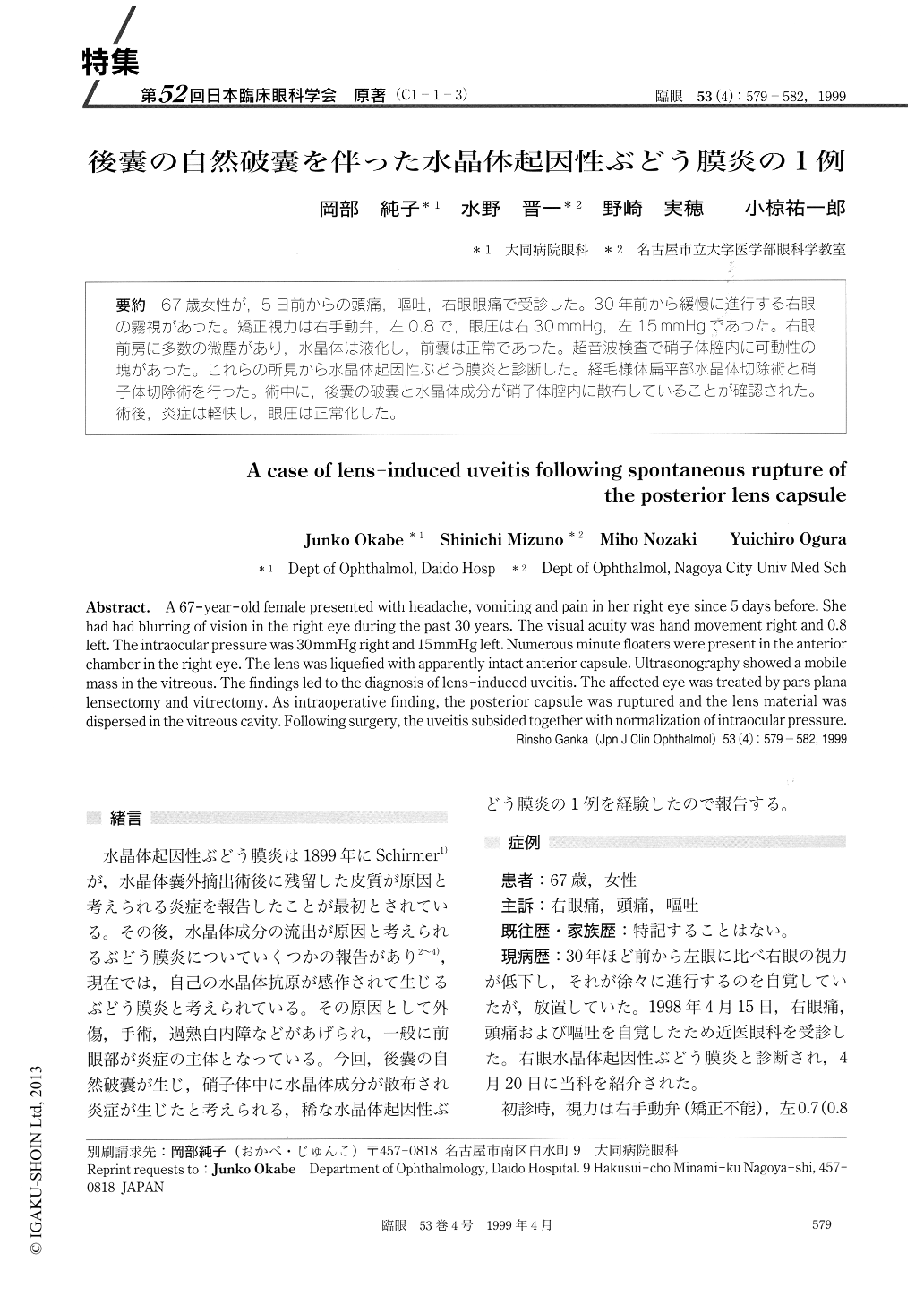
(C1-1-3) 67歳女性が,5日前からの頭痛,嘔吐,右眼眼痛で受診した。30年前から緩慢に進行する右眼の霧視があった。矯正視力は右手動弁,左0.8で,眼圧は右30mmHg,左15mmHgであった。右眼前房に多数の微塵があり,水晶体は液化し,前嚢は正常であった。超音波検査で硝子体腔内に可動性の塊があった。これらの所見から水晶体起因性ぶどう膜炎と診断した。経毛様体扁平部水晶体切除術と硝子体切除術を行った。術中に,後嚢の破嚢と水晶体成分が硝子体腔内に散布していることが確認された。術後,炎症は軽快し,眼圧は正常化した。
A 67-year-old female presented with headache, vomiting and pain in her right eye since 5 days before. She had had blurring of vision in the right eye during the past 30 years. The visual acuity was hand movement right and 0.8 left. The intraocular pressure was 30mmHg right and 15mmHg left. Numerous minute floaters were present in the anterior chamber in the right eye. The lens was liquefied with apparently intact anterior capsule. Ultrasonography showed a mobile mass in the vitreous. The findings led to the diagnosis of lens-induced uveitis. The affected eye was treated by pars plana lensectomy and vitrectomy. As intraoperative finding, the posterior capsule was ruptured and the lens material was dispersed in the vitreous cavity. Following surgery, the uveitis subsided together with normalization of intraocular pressure.
Copyright © 1999, Igaku-Shoin Ltd. All rights reserved.