Japanese
English
- 有料閲覧
- Abstract 文献概要
- 1ページ目 Look Inside
- 参考文献 Reference
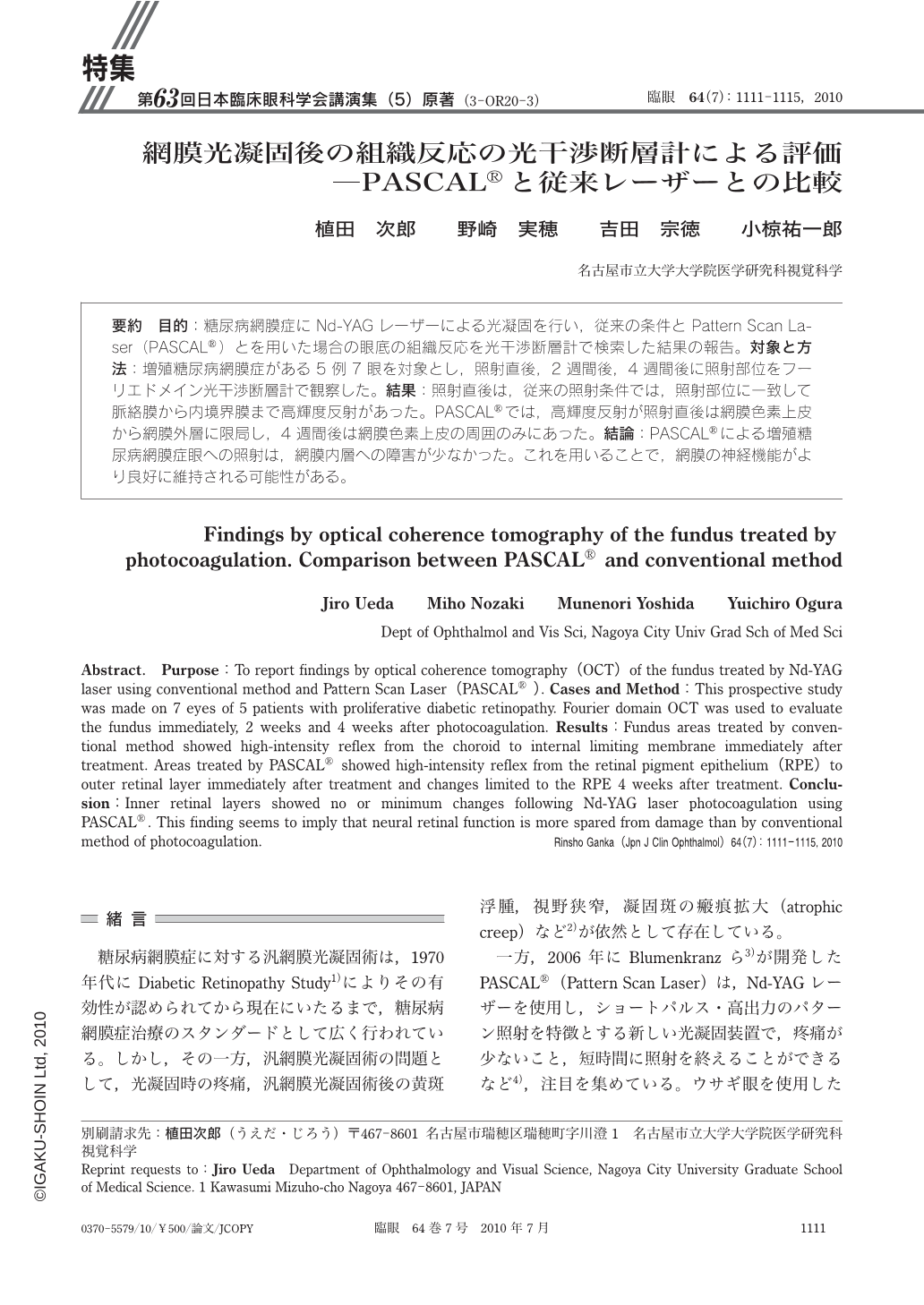
要約 目的:糖尿病網膜症にNd-YAGレーザーによる光凝固を行い,従来の条件とPattern Scan Laser(PASCAL®)とを用いた場合の眼底の組織反応を光干渉断層計で検索した結果の報告。対象と方法:増殖糖尿病網膜症がある5例7眼を対象とし,照射直後,2週間後,4週間後に照射部位をフーリエドメイン光干渉断層計で観察した。結果:照射直後は,従来の照射条件では,照射部位に一致して脈絡膜から内境界膜まで高輝度反射があった。PASCAL®では,高輝度反射が照射直後は網膜色素上皮から網膜外層に限局し,4週間後は網膜色素上皮の周囲のみにあった。結論:PASCAL®による増殖糖尿病網膜症眼への照射は,網膜内層への障害が少なかった。これを用いることで,網膜の神経機能がより良好に維持される可能性がある。
Abstract. Purpose:To report findings by optical coherence tomography(OCT)of the fundus treated by Nd-YAG laser using conventional method and Pattern Scan Laser(PASCAL®). Cases and Method:This prospective study was made on 7 eyes of 5 patients with proliferative diabetic retinopathy. Fourier domain OCT was used to evaluate the fundus immediately,2 weeks and 4 weeks after photocoagulation. Results:Fundus areas treated by conventional method showed high-intensity reflex from the choroid to internal limiting membrane immediately after treatment. Areas treated by PASCAL® showed high-intensity reflex from the retinal pigment epithelium(RPE)to outer retinal layer immediately after treatment and changes limited to the RPE 4 weeks after treatment. Conclusion:Inner retinal layers showed no or minimum changes following Nd-YAG laser photocoagulation using PASCAL®. This finding seems to imply that neural retinal function is more spared from damage than by conventional method of photocoagulation.

Copyright © 2010, Igaku-Shoin Ltd. All rights reserved.


