Japanese
English
- 有料閲覧
PPV購入案内
1,320円 (1,200円+税10%) こちらは、1397ページから1400ページの文献です。購入には医書.jp本会員登録が必要です。
会員登録完了後に改めて上記「購入サイトへ」を選択してください。
または「購入サイトへ」選択後に「購入ログイン」、「新規会員登録」の順に進んでください。
会員登録について詳しくはをご確認ください。
- Abstract 文献概要
- 1ページ目 Look Inside
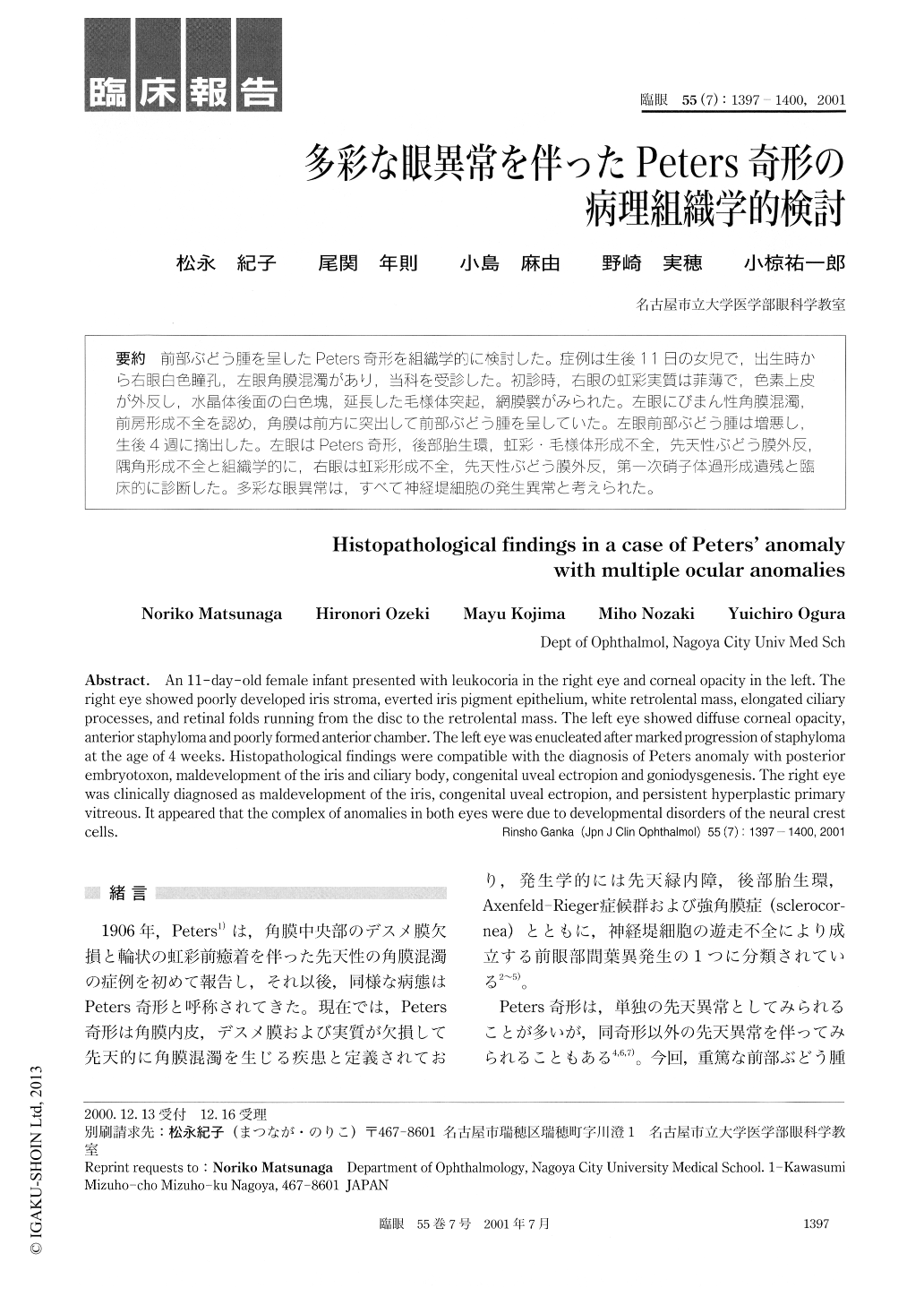
前部ぶどう腫を呈したPeters奇形を組織学的に検討した。症例は生後11日の女児で,出生時から右眼白色瞳孔,左眼角膜混濁があり,当科を受診した。初診時,右眼の虹彩実質は菲薄で,色素上皮が外反し,水晶体後面の白色塊,延長した毛様体突起,網膜襞がみられた。左眼にびまん性角膜混濁,前房形成不全を認め,角膜は前方に突出して前部ぶどう腫を呈していた。左眼前部ぶどう腫は増悪し,生後4週に摘出した。左眼はPeters奇形,後部胎生環,虹彩・毛様体形成不全,先天性ぶどう膜外反,隅角形成不全と組織学的に,右眼は虹彩形成不全,先天性ぶどう膜外反,第一次硝子体過形成遺残と臨床的に診断した。多彩な眼異常は,すべて神経堤細胞の発生異常と考えられた。
An 11-day-old female infant presented with leukocoria in the right eye and corneal opacity in the left. The right eye showed poorly developed iris stroma, everted iris pigment epithelium, white retrolental mass, elongated ciliary processes, and retinal folds running from the disc to the retrolental mass. The left eye showed diffuse corneal opacity,anterior staphyloma and poorly formed anterior chamber. The left eye was enucleated after marked progression of staphyloma at the age of 4 weeks. Histopathological findings were compatible with the diagnosis of Peters anomaly with posterior embryotoxon, maldevelopment of the iris and ciliary body, congenital uveal ectropion and goniodysgenesis. The right eye was clinically diagnosed as maldevelopment of the iris, congenital uveal ectropion, and persistent hyperplastic primary vitreous. It appeared that the complex of anomalies in both eyes were due to developmental disorders of the neural crest cells.
Copyright © 2001, Igaku-Shoin Ltd. All rights reserved.