Japanese
English
- 有料閲覧
PPV購入案内
1,320円 (1,200円+税10%) こちらは、173ページから176ページの文献です。購入には医書.jp本会員登録が必要です。
会員登録完了後に改めて上記「購入サイトへ」を選択してください。
または「購入サイトへ」選択後に「購入ログイン」、「新規会員登録」の順に進んでください。
会員登録について詳しくはをご確認ください。
- Abstract 文献概要
- 1ページ目 Look Inside
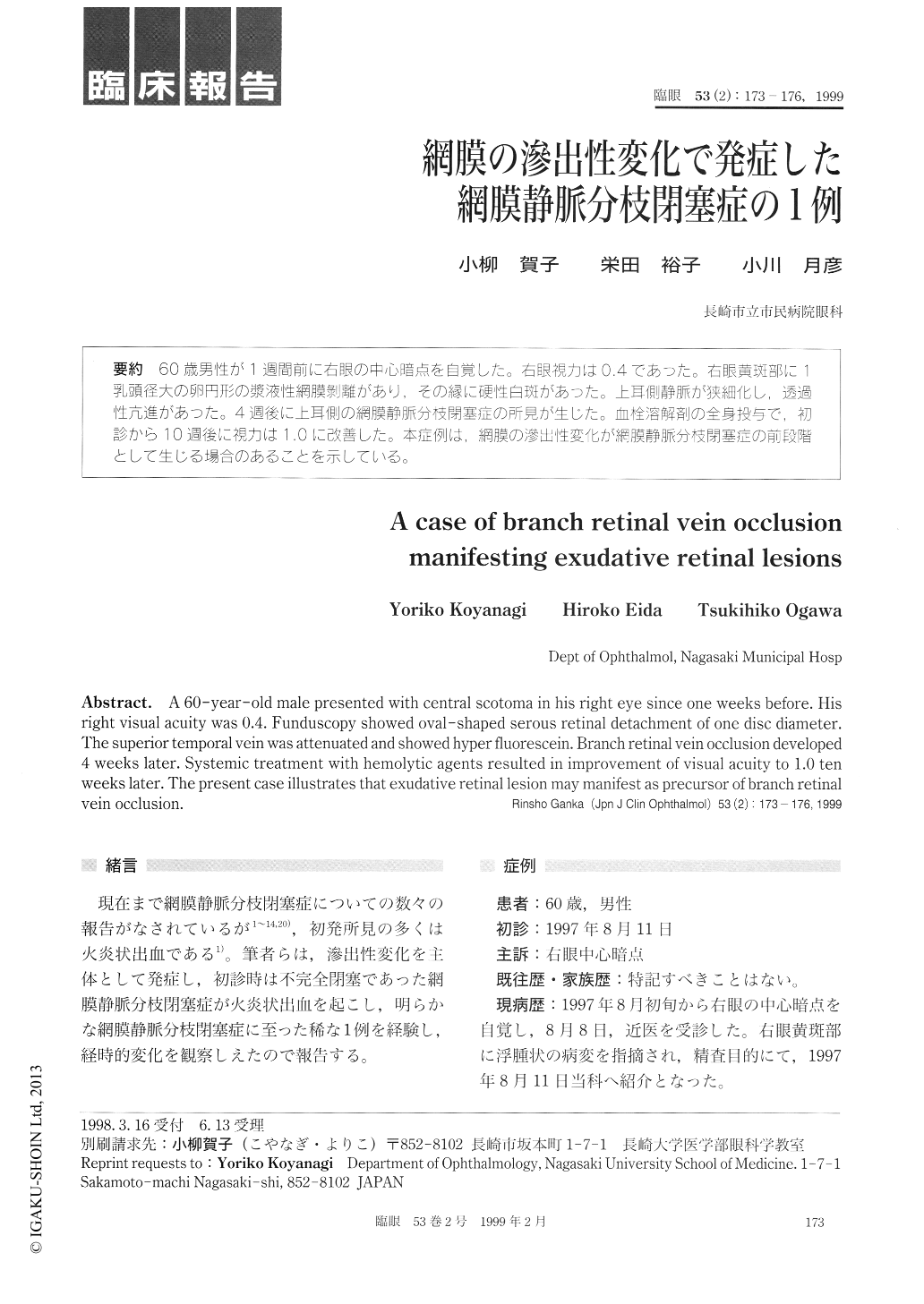
60歳男性が1週間前に右眼の中心暗点を自覚した。右眼視力は0.4であった。右眼黄斑部に1乳頭径大の卵円形の漿液性網膜剥離があり,その縁に硬性白斑があった。上耳側静脈が狭細化し,透過性亢進があった。4週後に上耳側の網膜静脈分枝閉塞症の所見が生じた。血栓溶解剤の全身投与で、初診から10週後に視力は1.0に改善した。本症例は,網膜の滲出性変化が網膜静脈分枝閉塞症の前段階として生じる場合のあることを示している。
A 60-year-old male presented with central scotoma in his right eye since one weeks before. His right visual acuity was 0.4. Funduscopy showed oval-shaped serous retinal detachment of one disc diameter. The superior temporal vein was attenuated and showed hyper fluorescein. Branch retinal vein occlusion developed 4 weeks later. Systemic treatment with hemolytic agents resulted in improvement of visual acuity to 1.0 ten weeks later. The present case illustrates that exudative retinal lesion may manifest as precursor of branch retinal vein occlusion.
Copyright © 1999, Igaku-Shoin Ltd. All rights reserved.