Japanese
English
- 有料閲覧
PPV購入案内
1,320円 (1,200円+税10%) こちらは、367ページから377ページの文献です。購入には医書.jp本会員登録が必要です。
会員登録完了後に改めて上記「購入サイトへ」を選択してください。
または「購入サイトへ」選択後に「購入ログイン」、「新規会員登録」の順に進んでください。
会員登録について詳しくはをご確認ください。
- Abstract 文献概要
- 1ページ目 Look Inside
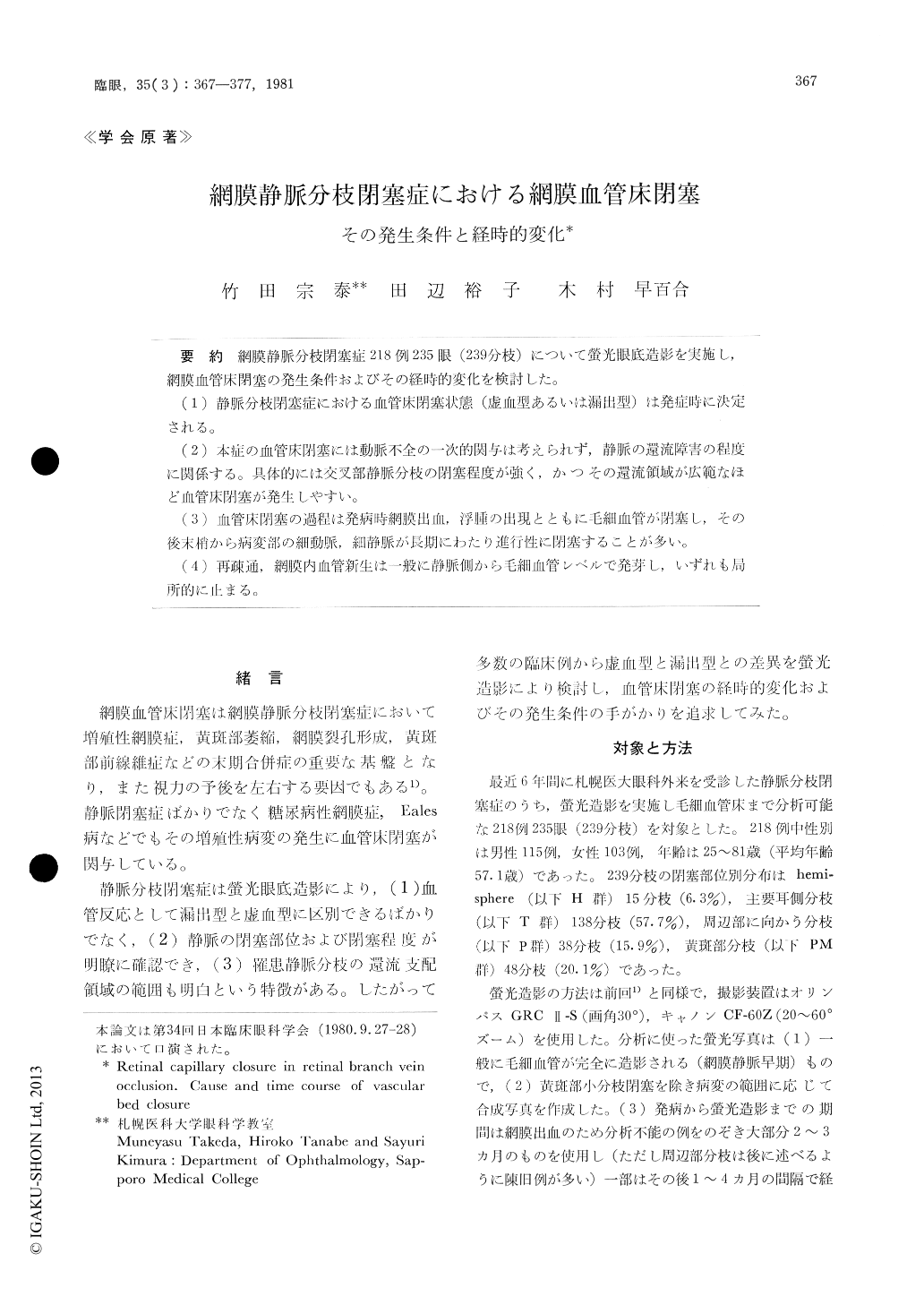
網膜静脈分枝閉塞症218例235眼(239分枝)について螢光眼底造影を実施し,網膜血管床閉塞の発生条件およびその経時的変化を検討した。
(1)静脈分枝閉塞症における血管床閉塞状態(虚血型あるいは漏出型)は発症時に決定される。
(2)本症の血管床閉塞には動脈不全の一次的関与は考えられず,静脈の還流障害の程度に関係する。具体的には交叉部静脈分枝の閉墓程度が強く,かつその還流領域が広範なほど血管床閉塞が発生しやすい。
(3)血管床閉塞の過程は発病時網膜出血,浮腫の出現とともに丘細血管が閉塞し,その後末梢から病変部の細動脈,細静脈が長期にわたり進行性に閉塞することが多い。
(4)再疎通,網膜内血管新生は一般に静脈側から毛細血管レベルで発芽し,いずれも局所的に止まる。
We evaluated the microvascular lesions and natural history of retinal branch vein occlusion in 235 eyes (239 sites) of 218 affected cases. A mon-tage technique of fluorescein fundus angiography was routinely used.
Retinal capillary nonperfusion was identified im-mediately or soon after onset of ischemic type of retinal branch vein occlusion. There after, closure of precapillary arterioles and postcapillary venules proceeded over the subsequent several months. Finally, these occlused venules or arterioles were barely identifiable as short stumps protruding from major retinal vessels.
Copyright © 1981, Igaku-Shoin Ltd. All rights reserved.