Japanese
English
- 有料閲覧
- Abstract 文献概要
- 1ページ目 Look Inside
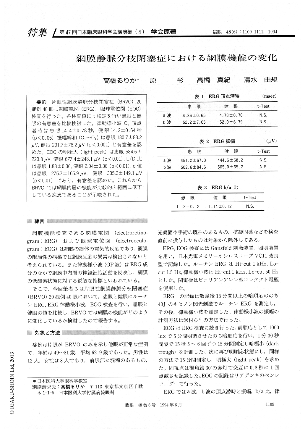
片眼性網膜静脈分枝閉塞症(BRVO)20症例40眼に網膜電図(ERG),眼球電位図(EOG)検査を行った。各検査値にt検定を行い患眼と健眼の有意差を比較検討した。律動様小波O1頂点潜時は患眼14.4±0.78秒,健眼14.2±0.64秒(p<0.05),振幅総和(O1〜O4)は患眼180.7±83.2μV,健眼231.7±78.2μV (p<0.001)と有意差を認めた。EOGの明極大(light peak)は患眼584.6±223.8μV,健眼677.4±248.1μV (p<0.01),L/D比は患眼1.83±0.36,健眼2.04±0.36(p<0.01),d値は患眼 275.7±165.9μV,健眼 335.2±149.1μV(p<0.01)であり,有意差を認めた。これらからBRVOでは網膜内層の機能が比較的広範囲に低下している疾患であることが示唆された。
We evaluated 20 patients with unilateral branch retinal vein occlusion using electroretinogram (ERG) and electrooculogram (EOG). The O1 peak time of oscillatory potentials was 14.4±0.78 msec in the affected and 14.2±0.64 msec in the unaffected fellow eyes (p<0.05). Summation of O1~O4 ampli-tudes was 180.7±83.2μV in the affected and 231.7±78.2μV in the fellow eyes (p<0.001). EOG light peak was 584.6 ±223.8μV in the affected and 677. 4±248.1μV in the fellow eyes (p<0.01). L/D ratio was 1.83±0.36 μV in the affected and 2.04±0.36 in the fellow eyes (p<0.01). D value was 275.7±165.9 μV in the affected and 335.2±149.1 μV in the fellow eyes (p<0.01). The findings indicate that the inner retinal layer function is diffusely impaired in branch retinal vein occlusion.

Copyright © 1994, Igaku-Shoin Ltd. All rights reserved.


