Japanese
English
- 有料閲覧
PPV購入案内
1,320円 (1,200円+税10%) こちらは、299ページから302ページの文献です。購入には医書.jp本会員登録が必要です。
会員登録完了後に改めて上記「購入サイトへ」を選択してください。
または「購入サイトへ」選択後に「購入ログイン」、「新規会員登録」の順に進んでください。
会員登録について詳しくはをご確認ください。
- Abstract 文献概要
- 1ページ目 Look Inside
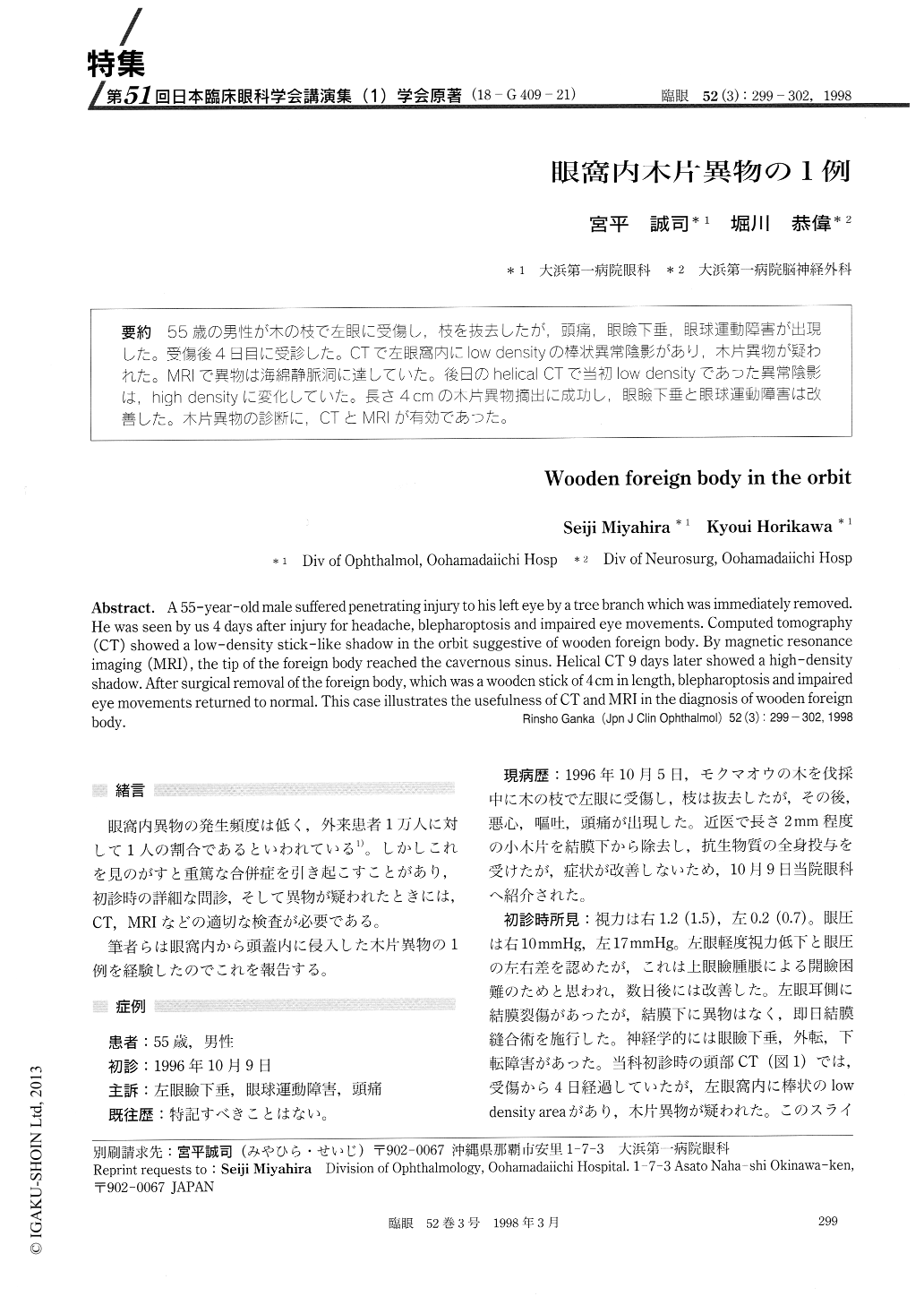
(18-G409-21) 55歳の男性が木の枝で左眼に受傷し,枝を抜去したが,頭痛,眼瞼下垂,眼球運動障害が出現した。受傷後4日目に受診した。CTで左眼窩内にlow densityの棒状異常陰影があり,木片異物が疑われた。MRIで異物は海綿静脈洞に達していた。後日のhelical CTで当初low densityであった異常陰影は,high densityに変化していた。長さ4cmの木片異物摘出に成功し,眼瞼下垂と眼球運動障害は改善した。木片異物の診断に,CTとMRIが有効であった。
A 55-year-old male suffered penetrating injury to his left eye by a tree branch which was immediately removed. He was seen by us 4 days after injury for headache, blepharoptosis and impaired eye movements. Computed tomography (CT) showed a low-density stick -like shadow in the orbit suggestive of wooden foreign body. By magnetic resonance imaging (MRI) , the tip of the foreign body reached the cavernous sinus. Helical CT 9 days later showed a high-density shadow. After surgical removal of the foreign body, which was a wooden stick of 4cm in length, blepharoptosis and impaired eye movements returned to normal. This case illustrates the usefulness of CT and MRI in the diagnosis of wooden foreign body.
Copyright © 1998, Igaku-Shoin Ltd. All rights reserved.