Japanese
English
- 有料閲覧
PPV購入案内
1,320円 (1,200円+税10%) こちらは、210ページから214ページの文献です。購入には医書.jp本会員登録が必要です。
会員登録完了後に改めて上記「購入サイトへ」を選択してください。
または「購入サイトへ」選択後に「購入ログイン」、「新規会員登録」の順に進んでください。
会員登録について詳しくはをご確認ください。
- Abstract 文献概要
- 1ページ目 Look Inside
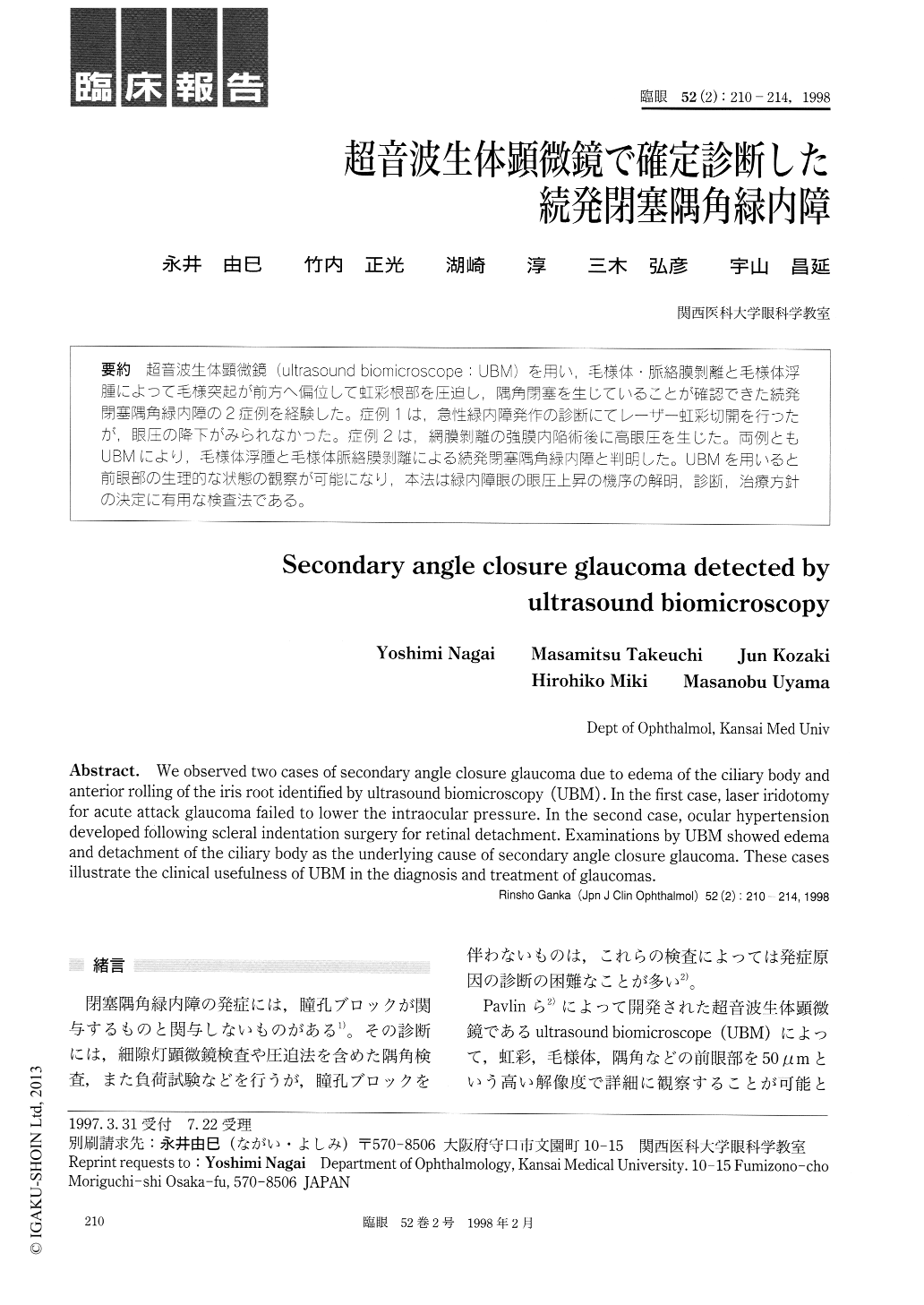
超音波生体顕微鏡(ultrasound biomicroscope:UBM)を用い,毛様体・脈絡膜剥離と毛様体浮腫によって毛様突起が前方へ偏位して虹彩根部を圧迫し,隅角閉塞を生じていることが確認できた続発閉塞隅角緑内障の2症例を経験した。症例1は,急性緑内障発作の診断にてレーザー虹彩切開を行ったが,眼圧の降下がみられなかった。症例2は,網膜剥離の強膜内陥術後に高眼圧を生じた。両例ともUBMにより,毛様体浮腫と毛様体脈絡膜剥離による続発閉塞隅角緑内障と判明した。UBMを用いると前眼部の生理的な状態の観察が可能になり,本法は緑内障眼の眼圧上昇の機序の解明,診断,治療方針の決定に有用な検査法である。
We observed two cases of secondary angle closure glaucoma due to edema of the ciliary body and anterior rolling of the iris root identified by ultrasound biomicroscopy (UBM). In the first case, laser iridotomy for acute attack glaucoma failed to lower the intraocular pressure. In the second case, ocular hypertension developed following scleral indentation surgery for retinal detachment. Examinations by UBM showed edema and detachment of the ciliary body as the underlying cause of secondary angle closure glaucoma. These cases illustrate the clinical usefulness of UBM in the diagnosis and treatment of glaucomas.
Copyright © 1998, Igaku-Shoin Ltd. All rights reserved.