Japanese
English
- 有料閲覧
PPV購入案内
1,320円 (1,200円+税10%) こちらは、209ページから215ページの文献です。購入には医書.jp本会員登録が必要です。
会員登録完了後に改めて上記「購入サイトへ」を選択してください。
または「購入サイトへ」選択後に「購入ログイン」、「新規会員登録」の順に進んでください。
会員登録について詳しくはをご確認ください。
- Abstract 文献概要
- 1ページ目 Look Inside
緒 言
網膜芽細胞腫の非腫瘤部の形態学的研究には,Teng等1),松尾等2),中尾等3)のものがあり,種種の未分化な構造やその他の異常構造の存在が報告されている。これらの発生要因については,腫瘍部から出た化学物質によるとする説と,発育上の未分化性を示すものであるとする説とがある。しかし,これまで非腫瘤部におけるこれらの未分化な構造や異常構造を,腫瘤部からの距離との関係について研究したものは見当らない。
今回,われわれは,網膜芽細胞腫の摘出眼の網膜を,乳頭部より鋸状縁まで連続的に追究し,上記の点を電子顕微鏡的に検討した結果,二,三の興味ある所見を得たので報告する。
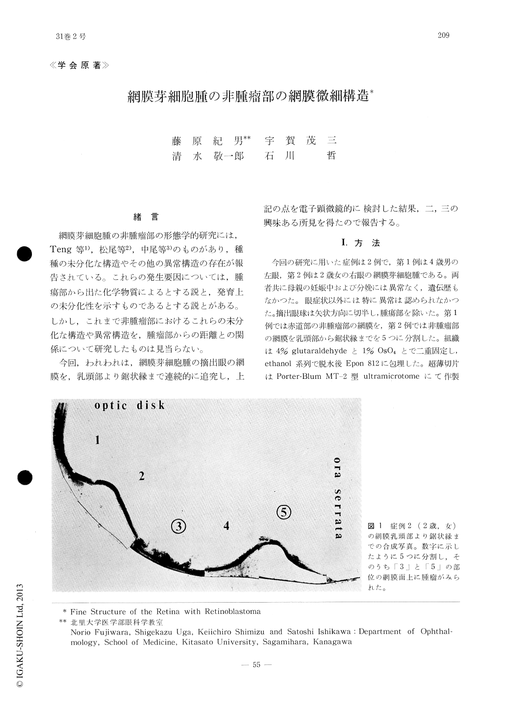
Structural differences between tumorous and non-tumorous areas of the retina, which was sampled in series from the peripapillary region to the ora serrata, were examined by electron microscopy in a case of retinoblastoma (2 year-old, female). The retinal portion with tumor mass on the inner limiting membrane contained (1) the nuclei of Müller cells which remained at the outer nuclear layer, (2) immature rod cells having cell organelles in the perinuclear cytoplasm, and (3) developing nerve processes which were characterized by vesicle formation at the tip of nerve processes and at the mar-ginal zone of the cytoplasm.
Copyright © 1977, Igaku-Shoin Ltd. All rights reserved.