Japanese
English
- 有料閲覧
PPV購入案内
1,320円 (1,200円+税10%) こちらは、861ページから865ページの文献です。購入には医書.jp本会員登録が必要です。
会員登録完了後に改めて上記「購入サイトへ」を選択してください。
または「購入サイトへ」選択後に「購入ログイン」、「新規会員登録」の順に進んでください。
会員登録について詳しくはをご確認ください。
- Abstract 文献概要
- 1ページ目 Look Inside
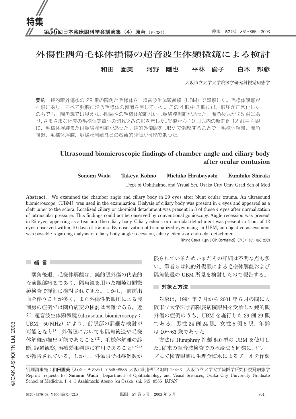
要約 鈍的眼外傷後の29眼の隅角と毛様体を,超音波生体顕微鏡(UBM)で観察した。毛様体解離が4眼にあり,すべて強膜に沿う毛様体の裂隙を呈していた。この4眼中3眼には,眼圧が正常化したのちでも,隅角鏡では見えない限局性の毛様体解離ないし脈絡膜剝離があった。隅角後退が25眼にあり,さまざまな程度の毛様体実質への切れ込みの形を示した。受傷から10日以内の新鮮例12眼中4眼に,毛様体浮腫または脈絡膜剝離があった。鈍的外傷眼をUBMで観察することで,毛様体解離,隅角後退,毛様体浮腫,脈絡膜剝離などの客観的評価が可能であった。
Abstract. We examined the chamber angle and ciliary body in 29 eyes after blunt ocular trauma. An ultrasound biomicroscope(UBM)was used in the examination. Dialysis of ciliary body was present in 4 eyes and appeared as a cleft inner to the sclera. Localized ciliary or choroidal detachment was present in 3 of these 4 eyes after normalization of intraocular pressure. This findings could not be observed by conventional gonioscopy. Angle recession was present in 25 eyes,appearing as a tear into the ciliary body. Ciliary edema or choroidal detachment was present in 4 out of 12 eyes observed within 10 days of trauma. By observation of traumatized eyes using an UBM,an objective assessment was possible regarding dialysis of ciliary body,angle recession,ciliary edema or choroidal detachment.
Copyright © 2003, Igaku-Shoin Ltd. All rights reserved.