Japanese
English
- 有料閲覧
- Abstract 文献概要
- 1ページ目 Look Inside
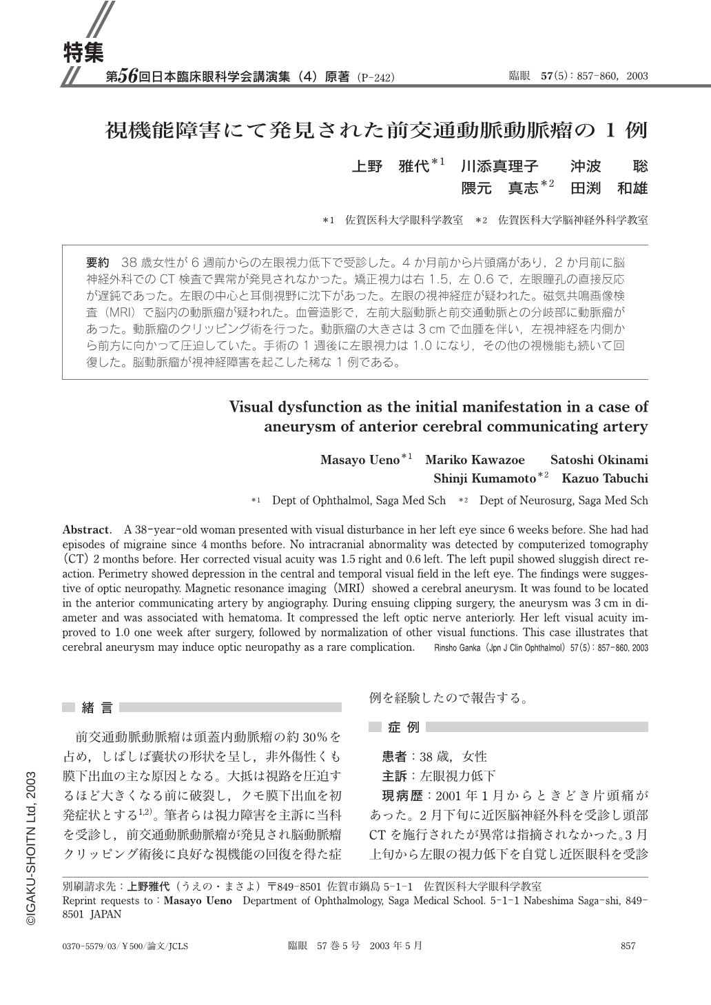
要約 38歳女性が6週前からの左眼視力低下で受診した。4か月前から片頭痛があり,2か月前に脳神経外科でのCT検査で異常が発見されなかった。矯正視力は右1.5,左0.6で,左眼瞳孔の直接反応が遅鈍であった。左眼の中心と耳側視野に沈下があった。左眼の視神経症が疑われた。磁気共鳴画像検査(MRI)で脳内の動脈瘤が疑われた。血管造影で,左前大脳動脈と前交通動脈との分岐部に動脈瘤があった。動脈瘤のクリッピング術を行った。動脈瘤の大きさは3cmで血腫を伴い,左視神経を内側から前方に向かって圧迫していた。手術の1週後に左眼視力は1.0になり,その他の視機能も続いて回復した。脳動脈瘤が視神経障害を起こした稀な1例である。
Abstract. A 38-year-old woman presented with visual disturbance in her left eye since 6 weeks before. She had had episodes of migraine since 4 months before. No intracranial abnormality was detected by computerized tomography(CT)2 months before. Her corrected visual acuity was 1.5 right and 0.6 left. The left pupil showed sluggish direct reaction. Perimetry showed depression in the central and temporal visual field in the left eye. The findings were suggestive of optic neuropathy. Magnetic resonance imaging(MRI)showed a cerebral aneurysm. It was found to be located in the anterior communicating artery by angiography. During ensuing clipping surgery,the aneurysm was 3 cm in diameter and was associated with hematoma. It compressed the left optic nerve anteriorly. Her left visual acuity improved to 1.0 one week after surgery,followed by normalization of other visual functions. This case illustrates that cerebral aneurysm may induce optic neuropathy as a rare complication.

Copyright © 2003, Igaku-Shoin Ltd. All rights reserved.


