Japanese
English
- 有料閲覧
PPV購入案内
1,320円 (1,200円+税10%) こちらは、1157ページから1162ページの文献です。購入には医書.jp本会員登録が必要です。
会員登録完了後に改めて上記「購入サイトへ」を選択してください。
または「購入サイトへ」選択後に「購入ログイン」、「新規会員登録」の順に進んでください。
会員登録について詳しくはをご確認ください。
- Abstract 文献概要
- 1ページ目 Look Inside
- 参考文献 Reference
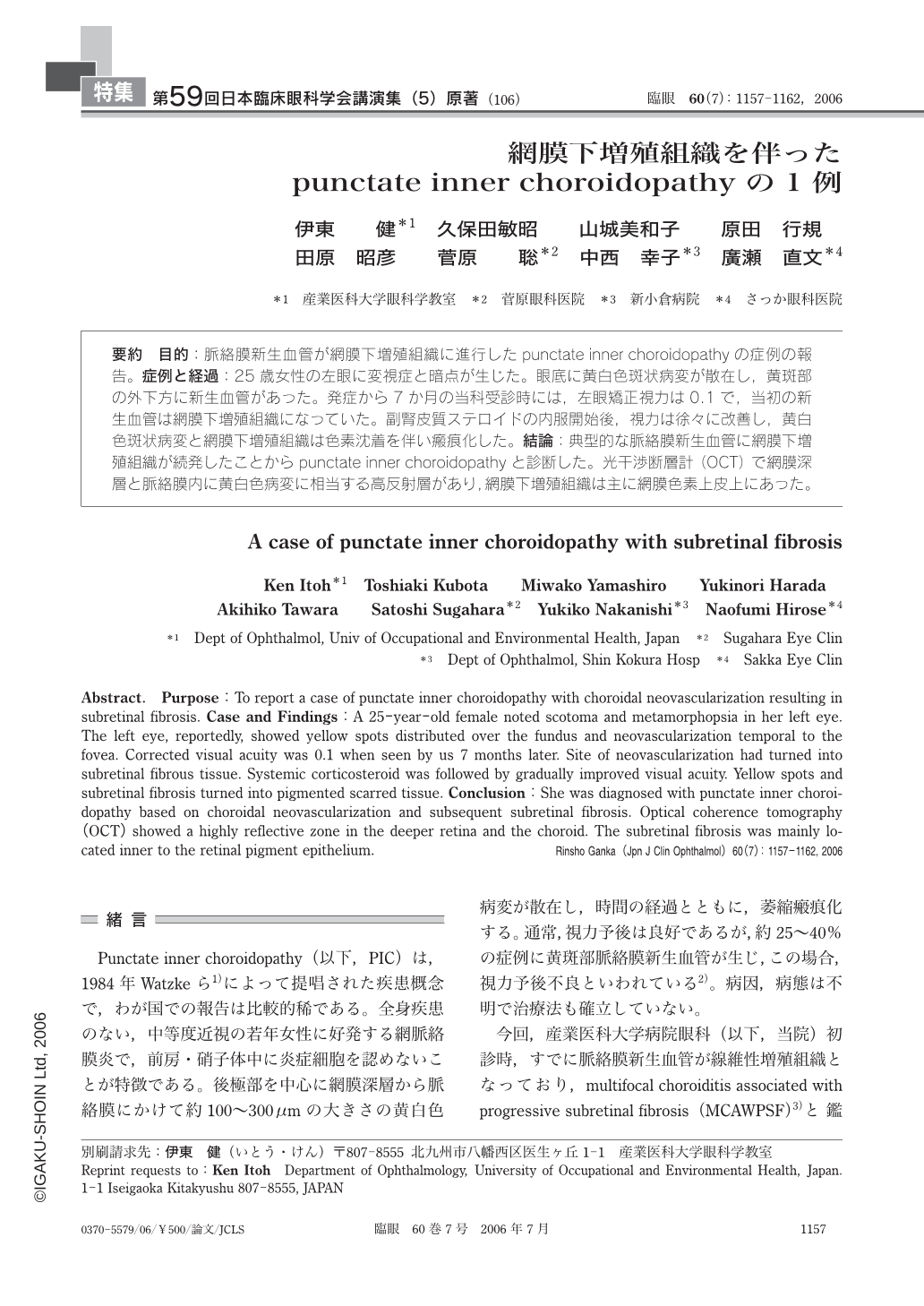
要約 目的:脈絡膜新生血管が網膜下増殖組織に進行したpunctate inner choroidopathyの症例の報告。症例と経過:25歳女性の左眼に変視症と暗点が生じた。眼底に黄白色斑状病変が散在し,黄斑部の外下方に新生血管があった。発症から7か月の当科受診時には,左眼矯正視力は0.1で,当初の新生血管は網膜下増殖組織になっていた。副腎皮質ステロイドの内服開始後,視力は徐々に改善し,黄白色斑状病変と網膜下増殖組織は色素沈着を伴い瘢痕化した。結論:典型的な脈絡膜新生血管に網膜下増殖組織が続発したことからpunctate inner choroidopathyと診断した。光干渉断層計(OCT)で網膜深層と脈絡膜内に黄白色病変に相当する高反射層があり,網膜下増殖組織は主に網膜色素上皮上にあった。
Abstract. Purpose:To report a case of punctate inner choroidopathy with choroidal neovascularization resulting in subretinal fibrosis. Case and Findings:A 25-year-old female noted scotoma and metamorphopsia in her left eye. The left eye,reportedly,showed yellow spots distributed over the fundus and neovascularization temporal to the fovea. Corrected visual acuity was 0.1 when seen by us 7 months later. Site of neovascularization had turned into subretinal fibrous tissue. Systemic corticosteroid was followed by gradually improved visual acuity. Yellow spots and subretinal fibrosis turned into pigmented scarred tissue. Conclusion:She was diagnosed with punctate inner choroidopathy based on choroidal neovascularization and subsequent subretinal fibrosis. Optical coherence tomography(OCT) showed a highly reflective zone in the deeper retina and the choroid. The subretinal fibrosis was mainly located inner to the retinal pigment epithelium.
Copyright © 2006, Igaku-Shoin Ltd. All rights reserved.